. 2022 Oct 29;23(21):13179.
doi: 10.3390/ijms232113179.

Apigenin Modulates AnxA6- and TNAP-Mediated Osteoblast Mineralization

Affiliations

Apigenin Modulates AnxA6- and TNAP-Mediated Osteoblast Mineralization

Joanna Mroczek et al. Int J Mol Sci. .

Abstract

Mineralization-competent cells like osteoblasts and chondrocytes release matrix vesicles (MVs) which accumulate Ca2+ and Pi, creating an optimal environment for apatite formation. The mineralization process requires the involvement of proteins, such as annexins (Anx) and tissue-nonspecific alkaline phosphatase (TNAP), as well as low molecular-weight compounds. Apigenin, a flavonoid compound, has been reported to affect bone metabolism, but there are doubts about its mechanism of action under physiological and pathological conditions. In this report, apigenin potency to modulate annexin A6 (AnxA6)- and TNAP-mediated osteoblast mineralization was explored using three cell lines: human fetal osteoblastic hFOB 1.19, human osteosarcoma Saos-2, and human coronary artery smooth muscle cells HCASMC. We compared the mineralization competence, the morphology and composition of minerals, and the protein distribution in control and apigenin-treated cells and vesicles. The mineralization ability was monitored by AR-S/CPC analysis, and TNAP activity was determined by ELISA assay. Apigenin affected the mineral structure and modulated TNAP activity depending on the concentration. We also observed increased mineralization in Saos-2 cells. Based on TEM-EDX, we found that apigenin influenced the mineral composition. This flavonoid also disturbed the intracellular distribution of AnxA6 and TNAP, especially blocking AnxA6 aggregation and TNAP attachment to the membrane, as examined by FM analysis of cells and TEM-gold analysis of vesicles. In summary, apigenin modulates the mineralization process by regulating AnxA6 and TNAP, as well as through various effects on normal and cancer bone tissues or atherosclerotic soft tissue.

Keywords: AnxA6; TNAP; apigenin; atherosclerosis; matrix vesicles; mineralization; osteoblast; osteosarcoma.


Conflict of interest statement

The authors declare no conflict of interest.

Figures

Figure 1
Figure 1
Morphology of hFOB 1.19 (A) and Saos-2 (B) cells. Hematoxylin–eosin staining of cells fixed in paraformaldehyde. Cells were observed under an optical microscope (magnification 400×).
Figure 2
Figure 2
The effect of apigenin on viability of hFOB 1.19 (squares), Saos-2 (circles) and HCAMSC (triangles) cells. The cells were incubated with different concentrations (µM) of apigenin for 7 days (hFOB 1.19 and Saos-2 cells) or 14 days (HCAMSC cells), then MTT assay was performed. Data are means ± S.E. of at least three independent experiments (* p < 0.05, ** p < 0.01 compared with cells without apigenin).
Figure 3
Figure 3
The effect of apigenin on mineralization of hFOB 1.19 (A) and Saos-2 (B) cells under resting conditions or in the presence of stimulators, AA and β-GP. Cells were incubated with different concentrations (µM) of apigenin for 7 days and then stained with AR-S and observed under an optical microscope (magnification 100×).
Figure 4
Figure 4
The effect of apigenin on the mineralization of hFOB 1.19 and Saos-2 cells. Quantitative analysis of calcium deposits in hFOB 1.19 (squares) and Saos-2 (circles) cells under resting conditions (open squares/circles) or in the presence of stimulators, AA, and β-GP (filled squares/circles) was carried out by staining with AR-S, de-staining with CPC, and absorbance measurements at λ 562 nm. The degree of mineralization was normalized to the relative number of viable cells. Data are means ± S.E. of at least three independent experiments (* p < 0.05, **/## p < 0.01 compared with resting/stimulated cells without apigenin).
Figure 5
Figure 5
The effect of apigenin on the TNAP activity of hFOB 1.19 (squares) and Saos-2 (circles) cells under resting conditions (open squares/circles) or in the presence of stimulators, AA, and β-GP (filled squares/circles). Cells were incubated with different concentrations (µM) of apigenin, and the TNAP activity assay (ALP Yellow pNPP Liquid Substrate System for ELISA) was performed in whole cell lysates after 7 days of culture. Absorbance at λ 405 nm was measured for 60 min with 15 s intervals at 37 °C. The TNAP activity values were normalized to the relative number of viable cells. Data are means ± S.E. of at least three independent experiments (* p < 0.05, **/## p < 0.01 compared with resting/stimulated cells without apigenin).
Figure 6
Figure 6
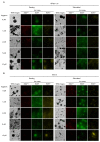
Co-localization of AnxA6 and TNAP during mineralization of hFOB 1.19 (A) and Saos-2 (B) cells under resting conditions or in the presence of stimulators, AA and β-GP. Cells were incubated with different concentrations (µM) of apigenin for 7 days, fixed and analyzed by fluorescent microscopy (magnification 630×). AnxA6 (green) was immunostained with anti-AnxA6 primary antibody conjugated with Alexa Fluor 488 secondary antibody. TNAP (red) was immunostained with anti-TNAP primary antibody conjugated with Alexa Fluor 594 secondary antibody. Sites of AnxA6 and TNAP co-localization are visible in yellow on merge images (arrows).
Figure 7
Figure 7
TEM images and MV chemical composition during the mineralization of hFOB 1.19 (A) and Saos-2 (B) cells under resting conditions or in the presence of stimulators, AA and β-GP. Cells were incubated with different concentrations (µM) of apigenin for 7 days, lysed, and analyzed by TEM-EDX (magnification 50,000×). Ion maps for Ca (red), p (green), and Cl (yellow) from EDX. Bar: 1 μm.
Figure 8
Figure 8
TEM images of the co-localization of AnxA6 and TNAP in MVs during the mineralization of hFOB 1.19 (A) and Saos-2 (B) cells under resting conditions or in the presence of stimulators, AA and β-GP. Cells were incubated with different concentrations (µM) of apigenin for 7 days, lysed, and analyzed by TEM (magnification 100,000×). Bar: 200 nm. Additional magnifications 300,000×. AnxA6 was labelled with anti-AnxA6 primary antibody conjugated with 10 nm colloidal gold secondary antibody. TNAP was labelled with anti-TNAP primary antibody conjugated with 5 nm colloidal gold secondary antibody. Sites of AnxA6 and TNAP co-localization are marked by squares for hFOB 1.19 cells and circles for Saos-2 cells.
Figure 9
Figure 9
Scheme of the effects of apigenin on the mineralization process. TNAP, attached to the membranes through a glycosylphosphatidylinositol (GPI) anchor, hydrolyzes inorganic pyrophosphate (PPi) into inorganic phosphate (Pi). AnxA6 may function as an inner and outer membrane protein as well as a transmembrane calcium ions (Ca2+) channel, whereas PiT may function as a Pi transporter. Apigenin stimulates (green arrow) hydroxyapatite (HA) formation from Ca2+ and Pi ions and inhibits (red block) AnxA6 and TNAP co-localization at the membrane of matrix vesicle (MV) secreted by the mineralizing cells to the extracellular matrix (ECM).

Similar articles

Cited by

References

    1. Aghajanian P., Mohan S. The art of building bone: Emerging role of chondrocyte-to-osteoblast transdifferentiation in endochondral ossification. Bone Res. 2018;6:19. doi: 10.1038/s41413-018-0021-z. - DOI - PMC - PubMed
    1. Anderson H.C. Molecular biology of matrix vesicles. Clin. Orthop. Relat. Res. 1995;314:266–280. doi: 10.1097/00003086-199505000-00034. - DOI - PubMed
    1. Anderson H.C. Matrix vesicles and calcification. Curr. Rheumatol. Rep. 2003;5:222–226. doi: 10.1007/s11926-003-0071-z. - DOI - PubMed
    1. Roy E Wuthier G.F.L. Matrix vesicles: Structure, composition, formation and function in calcification. Front. Biosci. 2011;16:2812–2902. doi: 10.2741/3887. - DOI - PubMed
    1. Thouverey C., Strzelecka-Kiliszek A., Balcerzak M., Buchet R., Pikula S. Matrix vesicles originate from apical membrane microvilli of mineralizing osteoblast-like Saos-2 cells. J. Cell. Biochem. 2009;106:127–138. doi: 10.1002/jcb.21992. - DOI - PubMed

MeSH terms

Grants and funding

This research was funded by a TRI-BIO-CHEM grant from the National Centre for Research and Development, POWER 3.2 Work implemented as a part of the Operational Programme Knowledge Education Development 2014–2020 and co-financed by the European Social Fund, Project No. POWR.03.02.00-00-I007/16-00 to J.M.; by an ERA-CVD/MICROEXPLORATION/4/2018 grant from the National Centre for Research and Development, Poland to S.P.; by the Hubert Curien Partnership Programme POLONIUM no PPN/BFR/2020/1/00056 for years 2021/2022 co-financed by the Polish National Agency for Academic Exchange and the Ministry of Foreign Affairs and International Development of France to A.S.-K. and by the statutory funds of the Nencki Institute of Experimental Biology, Polish Academy of Sciences.