. 2023 Aug;39(4):1715-1734.
doi: 10.1007/s10565-022-09776-4. Epub 2022 Nov 8.

Silica nanoparticles induce ovarian granulosa cell apoptosis via activation of the PERK-ATF4-CHOP-ERO1α pathway-mediated IP3R1-dependent calcium mobilization

Affiliations

Silica nanoparticles induce ovarian granulosa cell apoptosis via activation of the PERK-ATF4-CHOP-ERO1α pathway-mediated IP3R1-dependent calcium mobilization

Fenglei Chen et al. Cell Biol Toxicol. 2023 Aug.

Abstract

Ambient particulate matters (PMs) have adverse effects in human and animal female reproductive health. Silica nanoparticles (SNPs), as a major component of PMs, can induce follicular atresia via the promotion of ovarian granulosa cell apoptosis. However, the molecular mechanisms of apoptosis induced by SNPs are not very clear. This work focuses on revealing the mechanisms of ER stress on SNP-induced apoptosis. Our results showed that spherical Stöber SNPs (110 nm, 25.0 mg/kg b.w.) induced follicular atresia via the promotion of granulosa cell apoptosis by intratracheal instillation in vivo; meanwhile, SNPs decreased the viability and increase apoptosis in granulosa cells in vitro. SNPs were taken up and accumulated in the vesicles of granulosa cells. Additionally, our results found that SNPs increased calcium ion (Ca2+) concentration in granulosa cell cytoplasm. Furthermore, SNPs activated ER stress via an increase in the PERK and ATF6 pathway-related protein levels and IP3R1-dependent calcium mobilization via an increase in IP3R1 level. In addition, 4-PBA restored IP3R1-dependent calcium mobilization and decreased apoptosis via the inhibition of ER stress. The ATF4-C/EBP homologous protein (CHOP)-ER oxidoreductase 1 alpha (ERO1α) pathway regulated SNP-induced IP3R1-dependent calcium mobilization and cell apoptosis via ATF4, CHOP, and ERO1α depletion in ovarian granulosa cells. Herein, we demonstrate that ER stress cooperated in SNP-induced ovarian toxicity via activation of IP3R1-mediated calcium mobilization, leading to apoptosis, in which the PERK-ATF4-CHOP-ERO1α pathway plays an essential role in ovarian granulosa cells.

Keywords: Apoptosis; Calcium mobilization; ER stress; Follicular atresia; Ovarian granulosa cells; Silica nanoparticles.


Conflict of interest statement

Conflict of interest The authors declare no competing interests.

Figures

Fig. 1
Fig. 1
Effects of SNPs on follicular atresia. Aa Image of ovary tissue in the control. Ab Image of the selected area in Aa. Ba Image of ovary tissue after 12.5 mg/kg SNP treatment. Bb Image of the selected area in Ba. Ca Image of ovary tissue after 25.0 mg/kg SNP treatment. Cb Image of the selected area in Ca. Da Image of ovary tissue after 50.0 mg/kg SNP treatment. Db Image of the selected area in Da. Black solid arrow represents the healthy granulosa cells. Red solid arrow represents the apoptotic granulosa cells. E Image of ovary tis-sue after 50.0 mg/kg SNP treatment by TUNEL staining. F Image of the selected area in E. G Image of ovary tissue in the control by TUNEL staining. H Image of the selected area in G. White dotted arrow represents the apoptotic granulosa cells. Red dotted arrow represents the healthy granulosa cells. GCs, ovarian granulosa cells; O, oocyte
Fig. 2
Fig. 2
Cellular uptake of SNPs by TEM. A Representative image in the control. B Image of the selected blue area in A. C Image of the selected red area in B. D Representative image after 400 μg/mL SNP treatment for 24 h. E Image of the selected blue area in D. F Image of the selected red area in E. Blue arrow represents membranous vesicles without SNPs in ovarian granulosa cells. Red arrow represents membranous vesicles with SNPs in ovarian granulosa cells
Fig. 3
Fig. 3
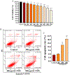
Determination of the cytotoxicity of SNPs in ovarian granulosa cells. A Measurement of cell viability. Granulosa cells were treated with or without SNPs at 100, 200, 300, 400, 500, 600, 800, 1000, and 1200 μg/mL concentrations for 24 h. B Detection of apoptosis. Granulosa cells were treated with or without SNPs at 200, 400, and 800 μg/mL concentrations for 24 h. C Quantification and statistical analysis in B. *p < 0.05, **p < 0.01, and ***p < 0.001 vs. control
Fig. 4
Fig. 4
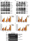
Activation of SNPs in ER stress. A Determination of GRP78, ATF6, p-PERK, p-eIF2α, ATF4, CHOP, and ERO1α levels. Granulosa cells were treated with or without SNPs at 200, 400, and 800 μg/mL concentrations for 12 h. B The gray analysis of GRP78, ATF6, p-PERK, and p-eIF2α in A. C The gray analysis of ATF4, CHOP, and EROla in A. D Determination of GRP78, ATF6, p-PERK, p-eIF2α, ATF4, CHOP, and ERO1α levels. Granulosa cells were treated with 400 μg/mL SNPs for 0–24 h. E The gray analysis of GRP78, ATF6, p-PERK, and p-eIF2α in D. F The gray analysis of ATF4, CHOP, and ERO1α in D. G Determination of Xbp-1 level by PCR. Granulosa cells were treated with 0–800 μg/mL SNPs, 10 μg/mL Tg, and 5 μg/mL Tm for 12 h. *p < 0.05, **p < 0.01, and ***p < 0.001 vs. control
Fig. 5
Fig. 5
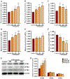
Effect of SNPs on IP3R-mediated calcium release. A–C Detection of [Ca2+]c by flow cytometry. Granulosa cells were treated with or without SNPs at 200, 400, and 800 μg/mL concentrations for 6 h (A), 12 h (B), and 24 h (C). D-F Determination of Ip3r1 (D), Ip3r2 (E), and Ip3r3 (F) Mrna levels. G Determination of IP3R1, IP3R2, and IP3R3 levels. Granulosa cells were treated with or without SNPs at 200, 400, and 800 μg/mL concentrations for 12 h. H The gray analysis of IP3R1, IP3R2, and IP3R3 in G. *p < 0.05, **p < 0.01, and ***p < 0.001 vs. control
Fig. 6
Fig. 6
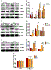
Inhibition of ER stress on SNP-induced cell apoptosis. A Measurement of cell viability. B Detection of [Ca2+]c by flow cytometry. C Detection of cell apoptosis. Granulosa cells were treated with or without SNPs at 200, 400, and 800 μg/mL concentrations for 24 h in the presence and absence of 0.5 mM 4-PBA. D Quantification and statistical analysis in C. E Determination of GRP78, p-PERK, p-eIF2α, ATF4, CHOP, and ERO1α levels. F The gray analysis of GRP78, p-PERK, and p-eIF2α in E. G The gray analysis of ATF4, CHOP, and ERO1α in E. H Determination of IP3R1, BCL-2, BAX, and cleaved caspase-3 levels. Granulosa cells were treated with 400 μg/mL of SNPs for 12 h in the presence and absence of 0.5 mM 4-PBA. I The gray analysis of IP3R1, the BAX/BCL-2 ratio, and cleaved caspase-3 levels in H. “−” represents that the cells were not treated with 4-PBA or SNPs, and “ + ” represents that the cells were treated with 4-PBA or SNPs. *p < 0.05, **p < 0.01, and ***p < 0.001 vs. control; #p < 0.05, ##p < 0.01, and ###p < 0.001 vs. SNP treatment
Fig. 6
Fig. 6
Inhibition of ER stress on SNP-induced cell apoptosis. A Measurement of cell viability. B Detection of [Ca2+]c by flow cytometry. C Detection of cell apoptosis. Granulosa cells were treated with or without SNPs at 200, 400, and 800 μg/mL concentrations for 24 h in the presence and absence of 0.5 mM 4-PBA. D Quantification and statistical analysis in C. E Determination of GRP78, p-PERK, p-eIF2α, ATF4, CHOP, and ERO1α levels. F The gray analysis of GRP78, p-PERK, and p-eIF2α in E. G The gray analysis of ATF4, CHOP, and ERO1α in E. H Determination of IP3R1, BCL-2, BAX, and cleaved caspase-3 levels. Granulosa cells were treated with 400 μg/mL of SNPs for 12 h in the presence and absence of 0.5 mM 4-PBA. I The gray analysis of IP3R1, the BAX/BCL-2 ratio, and cleaved caspase-3 levels in H. “−” represents that the cells were not treated with 4-PBA or SNPs, and “ + ” represents that the cells were treated with 4-PBA or SNPs. *p < 0.05, **p < 0.01, and ***p < 0.001 vs. control; #p < 0.05, ##p < 0.01, and ###p < 0.001 vs. SNP treatment
Fig. 7
Fig. 7
Depletion of IP3R1 on SNP-induced calcium release and apoptosis. A Determination of IP3R1, BCL-2, BAX, and cleaved caspase-3 levels. B, C, D The gray analysis of IP3R1 (B), the BAX/BCL-2 ratio (C), and cleaved caspase-3 (D) in A. E [Ca2+]c was monitored by flow cytometry. Ip3r1 lentiviruses were transduced into granulosa cells, and treated with 400 μg/mL SNPs for 12 h. F Detection of cell apoptosis. Ip3r1 lentiviruses were transduced into granulosa cells and treated with 400 μg/mL SNPs for 24 h. G Quantification and statistical analysis in F. *p < 0.05, **p < 0.01, and ***p < 0.001 vs. shNC treatment; #p < 0.05 and ##p < 0.01 vs. shNC + SNP treatment
Fig. 7
Fig. 7
Depletion of IP3R1 on SNP-induced calcium release and apoptosis. A Determination of IP3R1, BCL-2, BAX, and cleaved caspase-3 levels. B, C, D The gray analysis of IP3R1 (B), the BAX/BCL-2 ratio (C), and cleaved caspase-3 (D) in A. E [Ca2+]c was monitored by flow cytometry. Ip3r1 lentiviruses were transduced into granulosa cells, and treated with 400 μg/mL SNPs for 12 h. F Detection of cell apoptosis. Ip3r1 lentiviruses were transduced into granulosa cells and treated with 400 μg/mL SNPs for 24 h. G Quantification and statistical analysis in F. *p < 0.05, **p < 0.01, and ***p < 0.001 vs. shNC treatment; #p < 0.05 and ##p < 0.01 vs. shNC + SNP treatment
Fig. 8
Fig. 8
Inhibition of SNP-induced ER stress on IP3R1-mediated calcium release and apoptosis. A Determination of ATF4, CHOP, ERO1α, and IP3R1 levels. B The gray analysis of ATF4, CHOP, ERO1α, and IP3R1 in A. C Determination of CHOP, ERO1α, and IP3R1 levels. D The gray analysis of CHOP, ERO1α, and IP3R1 in C. E Determination of ERO1α and IP3R1 levels. F The gray analysis of ERO1α and IP3R1 in E. Atf4, Chop, and Ero1α lentiviruses were transduced into granulosa cells and treated with 400 μg/mL SNPs for 12 h, respectively. G Measurement of [Ca2+]c by flow cytometry. H Detection of cell apoptosis. Atf4, Chop, and Ero1α lentiviruses were transduced into granulosa cells and treated with 400 μg/mL SNPs for 24 h. I Quantification and statistical analysis in H. *p < 0.05, **p < 0.01, and ***p < 0.001 vs. shNC treatment; #p < 0.05 and ##p < 0.01 vs. shNC + SNP treatment
Fig. 8
Fig. 8
Inhibition of SNP-induced ER stress on IP3R1-mediated calcium release and apoptosis. A Determination of ATF4, CHOP, ERO1α, and IP3R1 levels. B The gray analysis of ATF4, CHOP, ERO1α, and IP3R1 in A. C Determination of CHOP, ERO1α, and IP3R1 levels. D The gray analysis of CHOP, ERO1α, and IP3R1 in C. E Determination of ERO1α and IP3R1 levels. F The gray analysis of ERO1α and IP3R1 in E. Atf4, Chop, and Ero1α lentiviruses were transduced into granulosa cells and treated with 400 μg/mL SNPs for 12 h, respectively. G Measurement of [Ca2+]c by flow cytometry. H Detection of cell apoptosis. Atf4, Chop, and Ero1α lentiviruses were transduced into granulosa cells and treated with 400 μg/mL SNPs for 24 h. I Quantification and statistical analysis in H. *p < 0.05, **p < 0.01, and ***p < 0.001 vs. shNC treatment; #p < 0.05 and ##p < 0.01 vs. shNC + SNP treatment
Fig. 9
Fig. 9
The diagram of the suggested mechanisms involved in SNP-induced apoptosis

Similar articles

Cited by

References

    1. Anelli T, Alessio M, Mezghrani A, Simmen T, Talamo F, Bachi A, et al. ERp44, a novel endoplasmic reticulum folding assistant of the thioredoxin family. EMBO J. 2002;21:835–44. - PMC - PubMed
    1. Averous J, Bruhat A, Jousse C, Carraro V, Thiel G, Fafournoux P. Induction of CHOP expression by amino acid limitation requires both ATF4 expression and ATF2 phosphorylation. J Biol Chem. 2004;279:5288–97. - PubMed
    1. Bitar A, Ahmad NM, Fessi H, Elaissari A. Silica-based nanoparticles for biomedical applications. Drug Discov Today. 2012;17:1147–54. - PubMed
    1. Che L, Yao H, Yang CL, Guo NJ, Huang J, Wu ZL, et al. Cyclooxygenase-2 modulates ER-mitochondria crosstalk to mediate superparamagnetic iron oxide nanoparticles induced hepatotoxicity: an in vitro and in vivo study. Nanotoxicology. 2020;14:162–80. - PubMed
    1. Chen F, Jin J, Hu J, Wang Y, Ma Z, Zhang J. Endoplasmic reticulum stress cooperates in silica nanoparticles-induced macrophage apoptosis via activation of CHOP-mediated apoptotic signaling pathway. Int J Mol Sci. 2019;20:5846. - PMC - PubMed

Publication types

LinkOut - more resources