. 2022 Oct 19:13:968776.
doi: 10.3389/fphar.2022.968776. eCollection 2022.

Potential predictive and therapeutic applications of small extracellular vesicles-derived circPARD3B in osteoarthritis

Affiliations

Potential predictive and therapeutic applications of small extracellular vesicles-derived circPARD3B in osteoarthritis

Zhiguo Lin et al. Front Pharmacol. .

Abstract

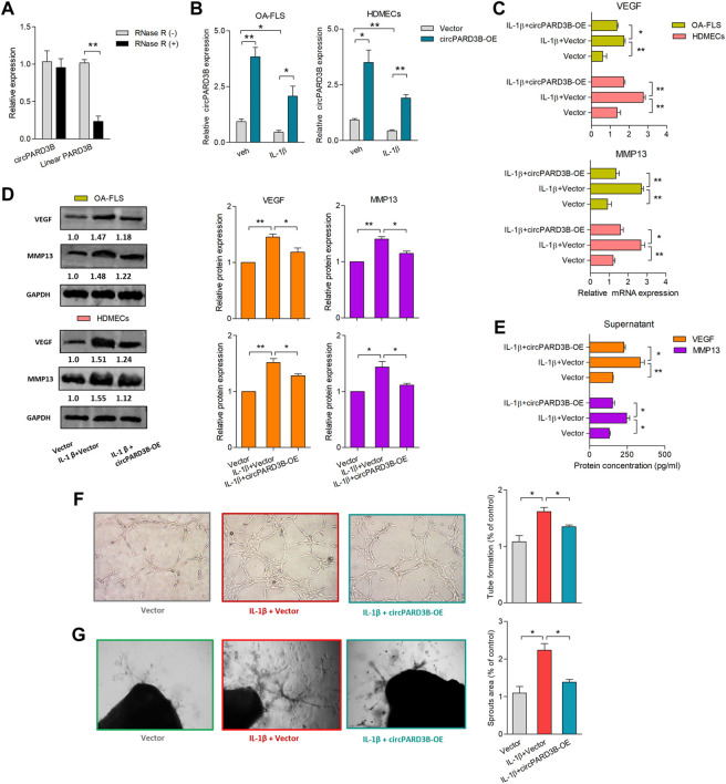
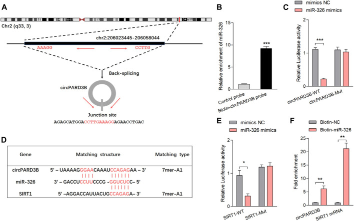
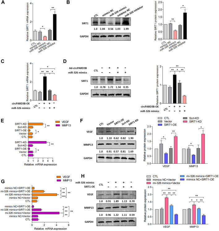
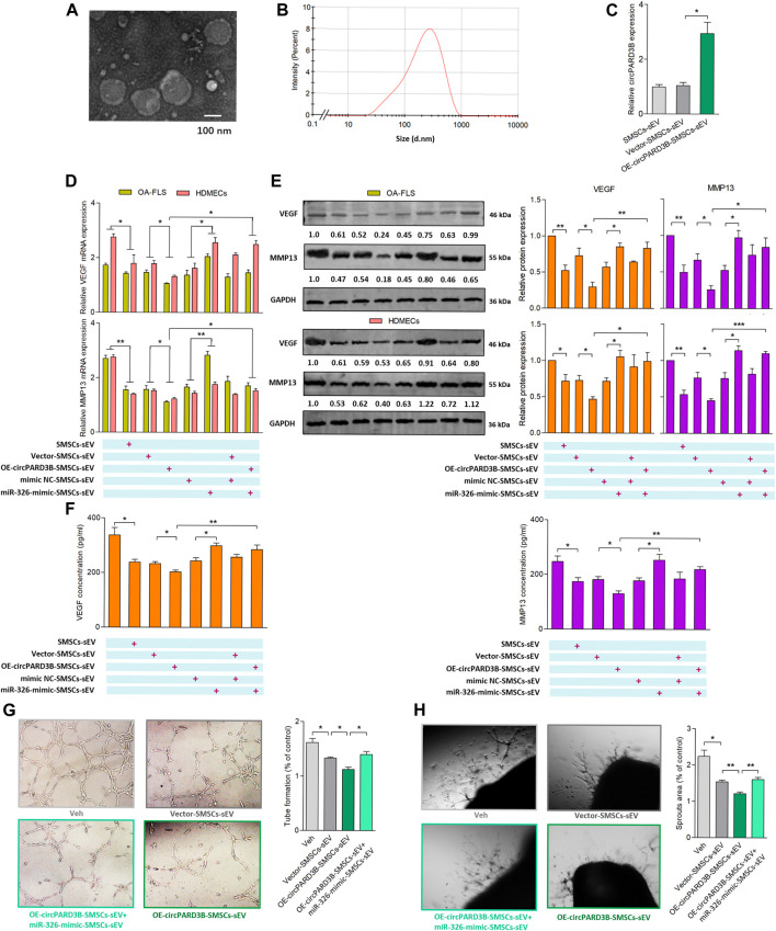
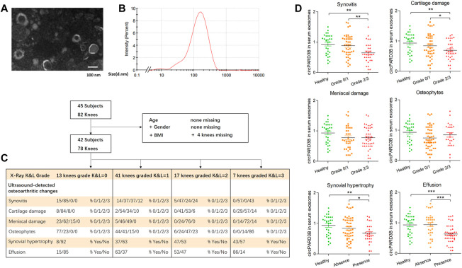
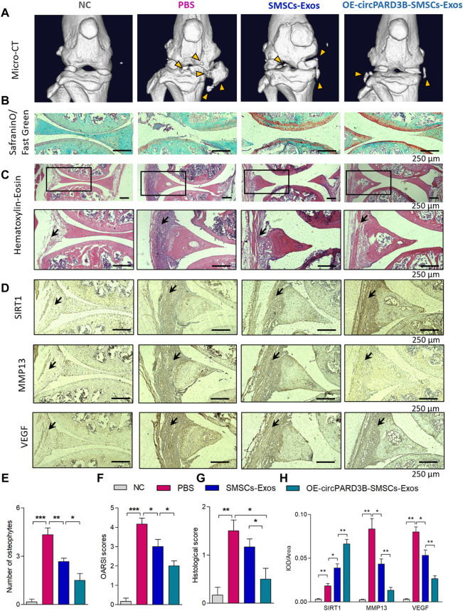
Background: Heterogeneous phenotypes that display distinct common characteristics of osteoarthritis (OA) are not well defined and will be helpful in identifying more customized therapeutic options for OA. Circular RNAs (circRNAs) have attracted more and more attention due to their role in the progression of OA. Investigating the role of circRNAs in the pathogenesis of OA will contribute to the phenotyping of OA and to individualized treatment. Methods: Small extracellular vesicles (sEV) were isolated from serum samples from patients with OA of different stages and sEV-derived circPARD3B was determined using RT-qPCR analysis. CircPARD3B expression in a stimulated coculture that included OA fibroblast-like synoviocytes (OA-FLS) as well as human dermal microvascular endothelial cells (HDMECs), plus the effects of circPARD3B on the expression of vascular endothelial growth factor (VEGF) long with angiogenic activity, were evaluated in vitro. Based on bioinformatics analysis and luciferase reporter assay (LRA), MiR-326 and sirtuin 1 (SIRT1) were found to be interactive partners of circPARD3B. Mesenchymal stem cells (SMSCs) overexpressing circPARD3B were constructed and SMSCs-derived sEV with overexpressed circPARD3B (OE-circPARD3B-SMSCs-sEV) were obtained to explore the effect of the intervention of circPARD3B combined with SMSCs-sEV-based therapy in vitro and in a OA model induced by collagenase in vivo. Results: Serum sEV-linked circPARD3B was indentified to be significantly decreased in the inflammatory phenotype of OA. Overexpression of circPARD3B was found to inhibit the expression of VEGF, as well as the angiogenesis induced by VEGF in a IL-1β stimulated the co-culture of OA-FLS as well as HDMECs. CircPARD3B is directly bound to miR-326. SIRT1 was considered a novel miR-326 target gene. OE-circPARD3B-SMSCs-sEV significantly reduced VEGF expression in coculture of OA-FLS and HDMECs. Injection of OE-circPARD3B-SMSCs-sEV could also reduce synovial VEGF; additionally, it could further ameliorate OA in the mouse model of OA in vivo. Conclusion: Serum sEV circPARD3B is a potential biomarker that enables the identification of the inflammatory phenotype of patients with OA. Correspondingly, intracellular transfer of circPARD3B through OE-circPARD3B-SMSCs-sEV could postpone disease progression through a functional module regulated angiogenesis of circPARD3B-miR-326-SIRT1, providing a novel therapeutic strategy for OA.

Keywords: SIRT1; VEGF; circPARD3B; osteoarthritis; small extracellular vesicles (sEV).


Conflict of interest statement

The authors declare that the research was conducted in the absence of any commercial or financial relationships that could be construed as a potential conflict of interest.

Figures

FIGURE 1
FIGURE 1
Overexpression of circPARD3B inhibited the production of VEGF and MMP13. mRNA (A–C) and protein (D) expression were respectively determined by qPCR and western blot analysis. (A) CircPARD3B’s resistance to Rnase R digestion (n = 3). (B) CircPARD3B’s expression in OA-FLS or HDMECs could be significantly downregulated by IL-1β induction, and then significantly elevated by circPARD3B overexpression (n = 3). (C–E) Role of circPARD3B overexpression in VEGF/MMP13 mRNA (C) as well as protein (D) expression in the OA-FLS or HDMECs co-culture, the concentration of VEGF in the supernatants of the co-culture (E) was significantly upregulated after induced by IL-1β, and then significantly downregulated via circPARD3B overexpression (n = 3). (F–G) Tube formation test (F)and ex vivo ARA of angiogenesis (G) proved the changes in the formation of capillary-like structure of HDMECs as well as microvessel sprouting with statistical significance based on the expression of VEGF in the supernatants of the OA-FLS and HDMECs co-culture (n = 3). Results are decribed with mean ± S.E.M. Veh = vehicle control. IL-1β = 10 ng/ml *p < 0.05, **p < 0.01.
FIGURE 2
FIGURE 2
CircPARD3B interacted with miR-326, further targeting SIRT1. (A) Characteristics of hsa_circ_0008172 (circPARD3B). (B) MiR-326 was pulled down and enriched with biotinylated circPARD3B probe (n = 3). (C) When co-transfected with wild circPARD3B as well as miR-326 mimics, the relative luc activity of circPARD3B was evidently suppressed (n = 3). (D) Schematic graph illustrating binding sites between circPARD3B and miR-326, miR-326 and SIRT1 mRNA. (E) LRA revealed that the luc activitiy of SIRT1 wild type reporter was decreased with statistical significance when transfected with miR-326 mimics in contrast with control reporter or mutated luciferase reporter (n = 3). (F) An enrichment of circPARD3B as well as SIRT1 mRNA in the pulled down sediments of 3′ end biotin-labeled miR-326 with statistical significance (n = 3). Results are described with mean ± S.E.M. CTL = control group. Scrl-KD = scramble siRNA knockdown. NC = negative control. *p < 0.05, **p < 0.01, ***p < 0.001.
FIGURE 3
FIGURE 3
Inhibition of VEGF/MMP13 expression via circPARD3B/miR-326/SIRT1 axis in vitro. mRNA and protein expression were respectively determined by qPCR and western blot analysis. (A,B) SIRT1 mRNA (A) as well as protein expression (B) were down- or up-regulated by miR-326 mimics or inhibitor with statistical significance (n = 3). (C,D) Co-effects of circPARD3B and miR-326 on the expression of SIRT1 in OA-FLS were assessed by the detection of SIRT1 mRNA (C) as well as protein (D) expression. SIRT1 expression were elevated or reduced by circPARD3B overexpression or miR-326 mimics with statistical significance in contrast to each control group. Besides, the downregulation of SIRT1 expression suppressed by miR-326 mimics was able to be evidently recovered by overexpressed circPARD3B, which means that the upregulated expression of SIRT1 induced by overexpressed circPARD3B was able to be inhibited by miR-326 mimics with statistical significance (n = 3). (E,F) The mRNA (E) as well as protein (F) expression of VEGF/MMP13 in OA-FLS were reduced or elevated by SIRT1 overexpression or knockdown with statistical significance (n = 3). (G,H) MiR-326 mimics upregulated VEGF/MMP13 mRNA (G) as well as protein (H) expression, and these increasement was able to be suppressed by SIRT1 overexpression with statistical significance (n = 3). Results are described with mean ± S.E.M. CTL = PBS control group. Scrl-KD = scramble siRNA knockdown. NC = negative control. *p < 0.05, **p < 0.01.
FIGURE 4
FIGURE 4
Evaluation of OE-circPARD3B-SMSCs-sEV’s anti-angiogenic effect on OA-FLS & HDMECs co-culture. SMSCs-sEV were confirmed by TEM (A) as well as DLS (B) as an average diameter of approximately 100 nm. mRNA (C,D) and protein (E) expression were respectively determined by qPCR and western blot analysis. (C) OE-circPARD3B-SMSCs-sEV carried enhanced expression of circPARD3B in contrast with those from Vector-SMSCs-sEV with statistical significance (n = 3). Under incubation of IL-1β, VEGF/MMP13 mRNA (D), as well as protein (E) expression in the OA-FLS and HDMECs co-culture and protein in the supernatants of the OA-FLS and HDMECs co-culture (F) were found to be downregulated by OE-circPARD3B-SMSCs-sEV, which was able to be inhibited by miR-326-mimic-SMSCs-sEV with statistical significance (n = 3). Changing trends of capillary-like structure formation in tube formation assay (G) as well as microvessel sprouting in ARA of angiogenesis (H) were in consistency with the changes of the expression of VEGF in supernatants (n = 3). Results are described with mean ± S.E.M. Veh = vehicle control *p < 0.05, **p < 0.01.
FIGURE 5
FIGURE 5
Serum-derived sEV circPARD3B as a potential biomarker for inflammatory phenotype of knee OA. Serum sEV were confirmed by TEM (A) and DLS (B). (C) Characteristic of OA patients under ultrasonography grading. (D) CircPARD3B levels in serum sEV were determined by qPCR. Correlations between serum sEV circPARD3B levels and sonographical grading characteristics of knee OA patients. Results are described with mean ± S.E.M *p < 0.05, **p < 0.01, ***p < 0.001.
FIGURE 6
FIGURE 6
Injection of OE-circPARD3B-SMSCs-sEV alleviate the progression of arthritis in collagenase-induced OA mouse model. (A) Representative micro-CT images of knee joints showing the abnormal growth of osteophytes (yellow arrow). (B) Representative images of Safranin O/Fast Green staining of knee cartilage from mice. (C) Representative images of HE staining analysis of knee joints from mice. After induced by OA, mice exhibited the mild inflammatory infiltration along with the synovial hyperplasia in the synovial tissues. Joint inflammation was reduced in the SMSCs-sEV group in contrast to the OA control group with statistical significance, especially in OE-circPARD3B-SMSCs-sEV group in contrast to BMSCs-sEV group. (D) IHC staining of SIRT1, VEGF and MMP13 were respectively demonstrated. We observed strengthened VEGF/MMP13 staining in synovial tissues of the OA control group in contrast to NC mice. OE-circPARD3B-SMSCs-sEV or SMSCs-sEV treated OA group revealed stregthened SIRT1 and weakened VEGF/MMP13 staining intensity in synovial tissues in contrast to the OA control mice, especially obvious in OE-circPARD3B-SMSCs-sEV group in contrast to that in the SMSCs-sEV group. (E) Quantification of images with 3D reconstruction by counting the number of osteophytes. (F) OARSI scoring to assess the histological changes of knee cartilage. (G) Histological assessment scores were shown. (H) Integrated optical density (IOD) values of immunostaining were shown in average. Scale bars = 250 µm. Data are described with mean ± S.E.M. n = 6; *p < 0.05, **p < 0.01, ***p < 0.001. NC = PBS treated normal control mice. OA control group = PBS treated OA group.
FIGURE 7
FIGURE 7
The mechanism of OE-circPARD3B-SMSCs-sEV therapy in OA. SMSCs stably overexpressing circPARD3B (OE-circPARD3B-SMSCs) were constructed after infected by the adenovirus. CircPARD3B derived from OE-circPARD3B-SMSCs might play a role as a sponge for the SIRT1-targeting miR-326. The downstream VEGF along with its conducted angiogenesis was then suppressed, with reduced expression of extracellular matrix (ECM) degrading enzyme MMP13 under local inflammatory microenvironment, such as IL-1β, ultimately inhibition of OA disease progression. Therefore, in contrast with the treatment of single SMSCs-sEV, the intracellular transfer of circPARD3B by OE-circPARD3B-SMSCs-sEV showed the better curative effect. Serum sEV circPARD3B level was found to be significantly downregulated in special phenotype OA patients with evident synovitis, suggesting that serum sEV circPARD3B might serve as a novel biomarker for the inflammatory phenotype of OA as well as an optimistic therapeutic effect of OE-circPARD3B-SMSCs-sEV towards this subgroup patients, indicating a potential novel therapeutic strategy for OA.

Similar articles

Cited by

References

    1. Altman R., Asch E., Bloch D., Bole G., Borenstein D., Brandt K., et al. (1986). Development of criteria for the classification and reporting of osteoarthritis. Classification of osteoarthritis of the knee. Diagnostic and Therapeutic Criteria Committee of the American Rheumatism Association. Arthritis Rheum. 29, 1039–1049. 10.1002/art.1780290816 - DOI - PubMed
    1. Alvaro-Gracia J. M., Zvaifler N. J., Firestein G. S. (1990). Cytokines in chronic inflammatory arthritis. V. Mutual antagonism between interferon-gamma and tumor necrosis factor-alpha on HLA-DR expression, proliferation, collagenase production, and granulocyte macrophage colony-stimulating factor production by rheumatoid arthritis synoviocytes. J. Clin. Invest. 86, 1790–1798. 10.1172/JCI114908 - DOI - PMC - PubMed
    1. Arden N., Richette P., Cooper C., BruyèRE O., Abadie E., Branco J., et al. (2015). Can we identify patients with high risk of osteoarthritis progression who will respond to treatment? A focus on biomarkers and frailty. Drugs Aging 32, 525–535. 10.1007/s40266-015-0276-7 - DOI - PMC - PubMed
    1. Batrakova E. V., Kim M. S. (2015). Using exosomes, naturally-equipped nanocarriers, for drug delivery. J. Control. Release 219, 396–405. 10.1016/j.jconrel.2015.07.030 - DOI - PMC - PubMed
    1. Berenbaum F. (2013). Osteoarthritis as an inflammatory disease (osteoarthritis is not osteoarthrosis!). Osteoarthr. Cartil. 21, 16–21. 10.1016/j.joca.2012.11.012 - DOI - PubMed

LinkOut - more resources