. 2023 Mar;71(3):524-540.
doi: 10.1002/glia.24291. Epub 2022 Nov 5.

Glial progenitor cells of the adult human white and grey matter are contextually distinct

Affiliations

Glial progenitor cells of the adult human white and grey matter are contextually distinct

Maria Joana Osorio et al. Glia. 2023 Mar.

Abstract

Genomic analyses have revealed heterogeneity among glial progenitor cells (GPCs), but the compartment selectivity of human GPCs (hGPCs) is unclear. Here, we asked if GPCs of human grey and white brain matter are distinct in their architecture and associated gene expression. RNA profiling of NG2-defined hGPCs derived from adult human neocortex and white matter differed in their expression of genes involved in Wnt, NOTCH, BMP and TGFβ signaling, suggesting compartment-selective biases in fate and self-renewal. White matter hGPCs over-expressed the BMP antagonists BAMBI and CHRDL1, suggesting their tonic suppression of astrocytic fate relative to cortical hGPCs, whose relative enrichment of cytoskeletal genes presaged their greater morphological complexity. In human glial chimeric mice, cortical hGPCs assumed larger and more complex morphologies than white matter hGPCs, and both were more complex than their mouse counterparts. These findings suggest that human grey and white matter GPCs comprise context-specific pools with distinct functional biases.

Keywords: NG2; glial heterogeneity; human glial progenitor cells; white and grey matter.


Conflict of interest statement

Dr. Goldman is also a part‐time employee and stock‐holder of Sana Biotechnology, a cell therapy company, and his lab receives sponsored research support from Sana. Maria Joana Osorio is currently employed at Sana. However, none of the work in this report was supported by Sana. No other authors have any known conflicts of interest in regard to this work.

Figures

FIGURE 1
FIGURE 1
hGPC density is conserved across adult human grey and white matter compartments. (a) The density of hGPCs was similar in the deep cortical GM (layers V/VI) and subcortical WM of the human temporal lobe. (b) The proportion of hGPCs among all cells was higher in the cortical GM than in the subcortical WM of the human temporal lobe. (c) The total cellular density was higher in the subcortical WM than in the cortical GM of the human temporal lobe. (d) Representative confocal images of NG2+ hGPCs (red) co‐labeled with nuclear OLIG2 (green) in the CTX and WM. Mean ± SEM. *p < .05, **p < .01 by unpaired t‐test. n = 4 samples. Scale: 20 μm. GM, grey matter; hGPC, human glial progenitor cell; WM, white matter
FIGURE 2
FIGURE 2
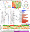
Adult hGPCs in the grey and white matter are transcriptionally distinct. Unsorted tissue and A2B5 selected hGPCs were obtained from WM and GM in four adult human patients and analyzed via microarray. (a) Principal component analysis of all samples illustrated separation of regional hGPCs. (b) Heatmap representation of differentially expressed CNS lineage markers in both WM and GM hGPCs, versus their respective unsorted tissue. (c) Intersection of differentially expressed genes (adjusted p < .05) in either tissue, versus their derived hGPCs or between WM or GM hGPCs. (d, e) Two curated networks were constructed of select ingenuity pathway analysis terms and their contributing genes for primarily glial (d), or cytoskeletal (e) terms. Circles represent genes, triangles IPA terms, and their relative size represents node degree. (f) Significance and activation Z‐scores of network IPA terms. Red indicates expression bias for grey matter GPCs; blue indicates white matter GPC bias. (g) Heatmap of the mean gene Z‐scores of all genes included in either network. Their presence in the glial or cytoskeletal network is indicated underneath. CNS, central nervous system; GM, grey matter; hGPC, human glial progenitor cell; WM, white matter
FIGURE 3
FIGURE 3
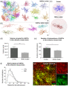
GM hGPCs are larger and more complex than WM hGPCs in adult glial chimeric mice. (a, b) Prototypic examples of 2D flat projections of 3D‐mapped hGPC morphologies in the CTX and callosum of the adult human glial chimeric mouse, as used for volumetric and branch analysis. (c) Representative 2D projections of individual, 3D‐mapped hGPCs, in both the CTX and CC of the adult human glial chimeric mouse used for Sholl analysis with superimposed 5 μm radius spheres. (d) hGPCs in the GM occupied larger volumes than those in the WM of human glial chimeric mice. (e) The total number of ramifications intersecting the Sholl's spheres was higher in cortical than in WM hGPCs. (f) Sholl analysis of individual hGPC morphologies in the CTX and CC of adult human glial chimeric mice revealed that cells within the CTX have increased branching at 15–35 μm distally from the cell body. (g) Representative confocal image of hGPCs in the mouse chimeric CTX. The human specific NG2+ is immunolabeled in red and overlapped with EGFP expression in a subset of cells, allowing a clear distinction of cell borders and hence reliable reconstruction of complete cells. Mean ± SEM. *p < .05, **p < .01, ***p < .001, ****p < .0001, by unpaired t‐test (d, e) and two‐way ANOVA with Bonferroni's post hoc (f). n = 4 mice. Scale: 20 μm. GM, grey matter; hGPC, human glial progenitor cell; WM, white matter
FIGURE 4
FIGURE 4
Human GPCs are larger and more complex than their mouse counterparts. (a) 2D projections of hGPCs and mGPCs 3D reconstructions in the CTX and WM, superimposed on the 5 μm radius spheres selected for the Sholl analysis. (b) hGPCs occupied larger volumes than mGPCs in the CTX but similar within the WM. mGPCs in the CTX occupied larger volumes than their counterparts in the underlying callosum. (c) hGPCs exhibited increased number of ramifications intersecting the Sholl's spheres than mGPCs, both in the CTX and callosum. mGPCs in the GM had higher number of ramifications intersecting the Sholl's spheres than mGPCs in the WM. (d) Sholl analysis of individual mGPCs showed that cells in the CTX were more complex than those in the WM at 15–35 μm distally from the cell body. (e, f) Sholl analysis revealed that hGPCs differ from mGPCs, with increased distal branching, both in the CTX (e) and WM (f). Mean ± SEM. *p < .05, **p < .01, ***p < .001 ****p < .0001 by unpaired t‐tests (b, c), and 2‐way ANOVA with Bonferroni's post hoc tests (d–f). n = 4 mice per group. Scale: 20 μm. GM, grey matter; hGPC, human glial progenitor cell; WM, white matter
FIGURE 5
FIGURE 5
BAMBI and CHRDL1 are differentially expressed by WM hGPCs relative to cortical hGPCs. In neonatally chimerized human glial chimeric mice sacrificed as adults, the BAMBI and CHRDL1 mRNA signal intensity was significantly higher in hGPCs resident within the callosal white matter (CC) than in their cortical counterparts (CTX). (a, b) In this analysis using RNAScope detection, the number of RNA dots was normalized to the area of the nucleus. Representative images are shown of cells in both compartments expressing BAMBI (a) and CHRDL1 (b) (red, BAMBI and CHRDL1; blue, DAPI); PDGFRA as a marker of hGPCs, green. *p < .05, **p < .01, by Mann–Whitney test. Scale 10 μm. CTX, cortex, CC, corpus callosum.
FIGURE 6
FIGURE 6
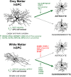
Key distinguishing features of GPCs of the human cortical grey matter and callosal white matter. Cortical hGPCs are larger and more complex than white matter hGPCs. Accordingly, they express genes involved in cytoskeletal function at higher levels compared to WM hGPCs. Whereas GM GPCs are enriched in pro‐astroglial transcripts involved in IL6 signaling, the expression of BMP antagonists by WM GPCs suggests an inhibition of astrogliogenesis at homeostatic baseline, favoring oligodendrocyte differentiation upon mobilization. GM, grey matter; hGPC, human glial progenitor cell; WM, white matter

Similar articles

Cited by

  • Survival of the fittest glia.
    Moser VA, Svendsen CN. Moser VA, et al. Nat Biotechnol. 2024 May;42(5):700-702. doi: 10.1038/s41587-023-01944-z. Nat Biotechnol. 2024. PMID: 37640947 No abstract available.

References

    1. Auvergne, R. M. , Sim, F. J. , Wang, S. , Chandler‐Militello, D. , Burch, J. , Al Fanek, Y. , Davis, D., Benraiss, A., Walter, K., Achanta, P., Johnson, M., Quinones‐Hinojosa, A., Natesan, N., Ford, H. L., & Goldman, S. A. (2013). Transcriptional differences between normal and glioma‐derived glial progenitor cells identify a core set of dysregulated genes. CELREP, 3(6), 2127–2141. 10.1016/j.celrep.2013.04.035 - DOI - PMC - PubMed
    1. Avilion, A. A., Nicolis, S. K., Pevny, L. H., Perez, L., Vivian, N., & Lovell‐Badge, R. (2003). Multipotent cell lineages in early mouse development depend on SOX2 function. Genes & Development, 17(1), 126–140. 10.1101/gad.224503 - DOI - PMC - PubMed
    1. Belachew, S. , Chittajallu, R. , Aguirre, A. A. , Yuan, X. , Kirby, M. , Anderson, S. , & Gallo, V. (2003). Postnatal NG2 proteoglycan‐expressing progenitor cells are intrinsically multipotent and generate functional neurons. The Journal of Cell Biology, 161(1), 169–186. 10.1083/jcb.200210110 - DOI - PMC - PubMed
    1. Benraiss, A. , Toner, M. J. , Xu, Q. , Bruel‐Jungerman, E. , Rogers, E. H. , Wang, F. , Economides, A. N., Davidson, B. L., Kageyama, R., Nedergaard, M., & Goldman, S. A. (2013). Sustained mobilization of endogenous neural progenitors delays disease progression in a transgenic model of Huntington's disease. Cell Stem Cell, 12(6), 787–799. 10.1016/j.stem.2013.04.014 - DOI - PMC - PubMed
    1. Bergles, D. E. , Roberts, J. D. , Somogyi, P. , & Jahr, C. E. (2000). Glutamatergic synapses on oligodendrocyte precursor cells in the hippocampus. Nature, 405(6783), 187–191. 10.1038/35012083 - DOI - PubMed

Publication types