. 2022 Nov 11;50(20):11682-11695.
doi: 10.1093/nar/gkac986.

A proto-telomere is elongated by telomerase in a shelterin-dependent manner in quiescent fission yeast cells

Affiliations

A proto-telomere is elongated by telomerase in a shelterin-dependent manner in quiescent fission yeast cells

Mélina Vaurs et al. Nucleic Acids Res. .

Abstract

Telomere elongation is coupled with genome replication, raising the question of the repair of short telomeres in post-mitotic cells. We investigated the fate of a telomere-repeat capped end that mimics a single short telomere in quiescent fission yeast cells. We show that telomerase is able to elongate this single short telomere during quiescence despite the binding of Ku to the proto-telomere. While Taz1 and Rap1 repress telomerase in vegetative cells, both shelterin proteins are required for efficient telomere extension in quiescent cells, underscoring a distinct mode of telomerase control. We further show that Rad3ATR and Tel1ATM are redundantly required for telomere elongation in quiescence through the phosphorylation of Ccq1 and that Rif1 and its associated-PP1 phosphatases negatively regulate telomerase activity by opposing Ccq1 phosphorylation. The distinct mode of telomerase regulation in quiescent fission yeast cells may be relevant to that in human stem and progenitor cells.


Figures

Figure 1.
Figure 1.
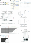
The 2R-48bp is extended by telomerase in quiescence. (A) The DNA sequence-specific endonuclease I-SceI is expressed under the control of a tetracycline-inducible promoter (TetR-controlled CaMV35S promoter) in a cassette that also expresses TetR. The addition of tetracycline (ahTet) induces I-SceI expression, which then cuts at site introduced into the genome. (B) The proto-telomere cassette is integrated downstream of the gal1+ gene, ∼47 kb from the right telomere of chromosome II (C2-R). The 2R-48 bp proto-telomere contains the ura4+ gene followed by 48 bp of telomere repeats (black triangles), a polylinker sequence (grey square), the I-SceI site (red triangle) and the hygromycin resistance marker (hph+), while the 0bp control lacks the telomere repeats. The S. pombe native telomere repeat tracts are indicated by gray triangles. Relative position of the restriction sites and probe used for Southern Blot analysis are indicated. (C) Schematic workflow of a typical quiescence experiment. Cells were grown in minimal medium (PMG + hygromycin) then shifted in nitrogen-depleted medium (MM-N) and maintained in quiescence for seven days. ahTet was added 8 h after nitrogen starvation to induce the proto-telomere. Samples were collected at different time points as indicated to perform genomic DNA extraction. (D, E) Genomic DNA from quiescent WT and ter1Δ cells was digested with ScaI and Southern blotted. Membrane was hybridized with ura4 probe to reveal the cut and uncut proto-telomere (2R-48 bp). D1, D3, D5 and D7 correspond to number of days for which cells were in G0 after ahTet addition before collecting and R indicates replicative sample collected before nitrogen-starvation. The membrane was also hybridized with abo1 probe used as a loading control. In D, the size (bp) of extended 2R-48bp is indicated. (F, G) Scatter plot representation of chromatin immunoprecipitation (ChIP) of Est1-V5 and Taz1-GFP in lig4Δ cells containing the 2R-48bp proto-telomere cassette. Quiescent cells were collected before I-SceI induction (D0) and 12H hours after induction with ahTet (D1). The immunoprecipitated DNA was analyzed by quantitative PCR with primers located nearby the cut (2R-48bp) and at 100 kb distance from the I-SceI site (control). The corrected % IP is the percentage of immunoprecipitated DNA of the target minus the control locus. Each dot corresponds to an individual experiment. Statistical comparisons were performed using an unpaired two-tailed t-test and a Mann–Whitney two-tailed test for Est1-V5 and Taz1-GFP ChIP, respectively (*** P-value < 0.005). (H, I) Schematic alignment of sequences obtained after 7 days in G0 (D7). For H the extended 2R-48 bp shown in panel D was gel extracted, cloned and sequenced. For I the shortened 2R-48 bp (the lower band of panel E) was gel extracted, cloned and sequenced. (J) After 7 days of nitrogen starvation in the presence of ahTet, G0 cells shown in panel D (2R-48 bp) were micromanipulated on rich YES-plates to allow exit from quiescence. Genomic DNA was extracted from seven individual clones, digested with ScaI and Southern blotted. Sample D7 of quiescence is loaded to compare the proto-telomere elongation to other samples. Membrane was hybridized with ura4 probe. Further extension of the 2R-48 bp was observed after cell cycle re-entry.
Figure 2.
Figure 2.
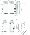
The NHEJ pathway and telomerase both process the proto-telomere in quiescence. (A–C) Genomic DNA from quiescent lig4Δ, pku70Δ and lig4Δ pku70Δ cells was digested with ScaI and Southern blotted. Membrane-bound DNA was hybridized with ura4 and abo1 probes. R (replicating cells), D0 (G0 before ahTet addition), D1, D3, D5, D7 and D10 (days in G0 after addition of ahTet) samples were collected. (D) Quantification of proto-telomere extension from panel A, B, C, and Figure 1D. Individual values are plotted from at least three different experiments, the colored and the black lines indicating the mean and the SEM, respectively. (E) Schematic alignment of sequences obtained from the shortest ura4+::2R-48 bp in pku70Δ cells after 7 days in G0 (D7). The lower band in panel B was gel extracted, cloned and sequenced. (F, G) Chromatin immunoprecipitation (ChIP) of Ku70-myc and Est1-V5 in lig4Δ cells containing the 2R-48bp proto-telomere cassette. Quiescent cells were collected before I-SceI induction (D0) and 12H hours after induction with ahTet (D1). The immunoprecipitated DNA was analyzed by quantitative PCR with primers located nearby the cut (2R-48 bp) and 100 kb away from the I-SceI site (control). The corrected % IP was plotted for each sample, and the values obtained in individual experiments are shown. Statistical comparisons were performed using an unpaired Mann–Whitney two-tailed test for Ku70-myc and Est1-V5ChIP (* P-value < 0.05 and *** P-value < 0.005).
Figure 3.
Figure 3.
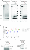
Rad3ATR controls telomerase activity through Ccq1 phosphorylation. (A–E) Genomic DNA from quiescent ccq1-T93A lig4Δ, tel1-kdΔ lig4Δ, rad3-kdΔ lig4Δ , rad3-kdΔ tel1-kdΔ lig4Δ and mre11Δ lig4Δ cells was digested with ScaI and Southern blotted. Membrane was hybridized with ura4 probe. R (replicating cells), D0 (G0 before ahTet addition), D1, D3, D5 and D7 (days in G0 after addition of Tc) samples were collected.
Figure 4.
Figure 4.
Taz1 and Rap1 positively regulate telomerase activity in quiescent cells. (A-B,D-E) Genomic DNA from quiescent taz1Δ lig4Δ , rap1Δ lig4Δ, rap1-I655R lig4Δ and tpz1-I200R lig4Δ cells was digested with ScaI and Southern blotted. Membrane was hybridized with ura4 probe. R (replicating cells), D0 (G0 before ahTet addition), D1, D3, D5 and D7 (days in G0 after addition of ahTet) samples were collected. (C) Analysis of the 2R-48bp extension in taz1Δ and rap1Δ vegetative cells after ahTet addition. Genomic DNA was extracted, digested with ScaI and Southern blotted. Membrane was hybridized with ura4 probe. (F) Ccq1-Flag ChIP in WT, rap1Δ and taz1Δ lig4Δ cells containing the 2R-48 bp proto-telomere cassette. Quiescent cells were collected before (D0) and 12H hours after (D1) I-SceI induction with ahTet. The immunoprecipitated DNA was analyzed by quantitative PCR with primers located nearby the cut (2R-48 bp) and 100 kb away from the I-SceI site (control). The corrected % IP was plotted with the values obtained in individual experiments shown for each sample. Statistical comparisons were performed using an unpaired Mann–Whitney two-tailed test to compare D0 and D1 of Ccq1-flag in WT and an unpaired two-tailed t-test for the others ** P-value < 0.01 and *** P-value < 0.005).
Figure 5.
Figure 5.
Stn1 and Rif1 negatively regulate telomerase activity in quiescent cells. (A–C) Genomic DNA from quiescent snt1-226 lig4Δ, rif1Δ lig4Δ, and rif1-PP1 lig4Δ cells was digested with ScaI and Southern blotted. Membrane was hybridized with ura4 probe. R (replicating cells), D0 (G0 before ahTet addition), D1, D3, D5 and D7 (days in G0 after addition of ahTet) samples were collected. (D) Quantification of proto-telomere extension shown in panel B, C and Figure 2A. Individual values are plotted from at least 3 different experiments, the colored and the black lines indicating the mean and the SEM, respectively. (E) The phosphorylation of Ccq1 from indicated strains carrying the 2R-48bp was assessed by Western blot. TCA extraction was performed from vegetative cells before nitrogen starvation and immunoblotted with anti-flag antibody. The slow-migrating band corresponds to phosphorylated Ccq1. Ponceau red staining was used as loading control. (F) The phosphorylation of Ccq1 from indicated strains carrying the 2R-48 bp was assessed by western blot. TCA extraction was performed from cell samples maintained one day in quiescence with addition of ahTet to induce the I-SceI cut.
Figure 6.
Figure 6.
Control of telomerase activity in vegetative and quiescent cells. (A) Telomere processing in quiescent cells. A short telomere is either recognized by Ku complex that will promote NHEJ or extended by telomerase. NHEJ is the most proficient pathway in G0 cells, however a telomeric seed is able to promote telomerase recruitment. (B) Telomerase activation in quiescent cells. Taz1 and Rap1 are both required to assemble shelterin via their bridging function to promote telomerase recruitment that requires the Rad3ATR/Tel1ATM-dependent phosphorylation of Ccq1. (C) Telomerase inhibition in quiescent cells. Inhibition of telomerase relies on the dephosphorylation of Ccq1 through Rif1-associated PP1 phosphatases and on the concomitant sumolylation of Tpz1 that promotes Stn1-Ten1 recruitment and synthesis of the complementary strand. Ku binding to the extended telomere is also thought to inhibit telomerase action. (D) Telomerase activation in cycling cells. Replication stress at short telomeres generates robust substrates, such as reversed forks, for telomerase. RPA-coated ssDNA promotes the Rad3ATR/Tel1ATM-dependent phosphorylation of Ccq1 which then recruits telomerase independently of Taz1-Rap1 shelterin assembly.

Similar articles

Cited by

References

    1. Lange T.de Shelterin-mediated telomere protection. Annu. Rev. Genet. 2018; 52:223–247. - PubMed
    1. Higa M., Fujita M., Yoshida K.. DNA replication origins and fork progression at mammalian telomeres. Genes (Basel). 2017; 8:112. - PMC - PubMed
    1. Maestroni L., Matmati S., Coulon S.. Solving the telomere replication problem. Genes. 2017; 8:55. - PMC - PubMed
    1. Taddei A., Gasser S.M.. Structure and function in the budding yeast nucleus. Genetics. 2012; 192:107–129. - PMC - PubMed
    1. Lingner J., Cooper J., Cech T.. Telomerase and DNA end replication: no longer a lagging strand problem?. Science. 1995; 269:1533–1534. - PubMed

Publication types

MeSH terms