. 2021 Oct 26;31(44):2009946.
doi: 10.1002/adfm.202009946. Epub 2021 Mar 26.

Single-Cell Microgels for Diagnostics and Therapeutics

Affiliations

Single-Cell Microgels for Diagnostics and Therapeutics

Ryan Dubay et al. Adv Funct Mater. .

Abstract

Cell encapsulation within hydrogel droplets is transforming what is feasible in multiple fields of biomedical science such as tissue engineering and regenerative medicine, in vitro modeling, and cell-based therapies. Recent advances have allowed researchers to miniaturize material encapsulation complexes down to single-cell scales, where each complex, termed a single-cell microgel, contains only one cell surrounded by a hydrogel matrix while remaining <100 μm in size. With this achievement, studies requiring single-cell resolution are now possible, similar to those done using liquid droplet encapsulation. Of particular note, applications involving long-term in vitro cultures, modular bioinks, high-throughput screenings, and formation of 3D cellular microenvironments can be tuned independently to suit the needs of individual cells and experimental goals. In this progress report, an overview of established materials and techniques used to fabricate single-cell microgels, as well as insight into potential alternatives is provided. This focused review is concluded by discussing applications that have already benefited from single-cell microgel technologies, as well as prospective applications on the cusp of achieving important new capabilities.

Keywords: 3D cell culture; cell-based therapies; hydrogels; regenerative medicine; single-cell analysis.


Conflict of interest statement

Conflict of Interest The authors declare no conflict of interest.

Figures

Figure 1.
Figure 1.
A) Influence of microgel size on physiological response and pharmacokinetics.[21] Cell diameter, Dcell =12 μm, and diffusion coefficient D = 10 μm2 s−1, were used. B) Components of single-cell microgels, with tunable characteristics to suit application needs. Figure (A) was reproduced with permission.[21] Copyright 2018, Elsevier.
Figure 2.
Figure 2.
A) Schematic of cell encapsulation using conventional (upper panel) and cell surface coating (lower panel) methods for crosslinking alginate microgels. B) Fraction of alginate microgels containing MSCs and OP9 cells via using conventional (Direct) encapsulation, conventional encapsulation followed by FACS, and cell surface pre-coating. E is theoretical yield for conventional encapsulation. C) Confocal image of encapsulated MSC (green, alginate; red, actin; blue, nucleus), scale bar denotes 10 μm.[46] D) Schematic of CLEX (competitive ligand exchange crosslinking). E) Image of microfluidic device used to fabricate cell-laden microgels using CLEX process, where numbers correspond to solutions in (F). F) Fluorescently labeled cell-free alginate microgels using CLEX (left) and release of Ca2+ from CaEDTA with acidic carrier fluid (right). CLEX microgels are more uniformly stained as a result of the gradual release of Ca2+, compared to non-uniform distribution as a result of rapid gelation. Alg., fluorescent alginate; HOAc, acetic acid; Surf., surfactant.[38] Images (A), (C), and figure (B) are reproduced with permission.[46] Copyright 2016, Springer Nature. Images (D–F) are reproduced with permission.[38] Copyright 2016, Royal Society of Chemistry.
Figure 3.
Figure 3.
A) Generation of agarose-based microgels. From left to right, images of initial droplet generation event for cell encapsulation, droplet mixer, first droplet splitter, and third droplet splitter. B) Normalized droplet diameter as a result of N splitting events. Images in (A) are reproduced with permission.[65] Copyright 2019, Royal Society of Chemistry.
Figure 4.
Figure 4.
A,B) Phase contrast images of acinus growth within a Matrigel microgel over time starting from a single cell (Day 1). White dashed lines indicate microgel periphery, for clarity. C) Fluorescent staining of actin (red) and nuclei (blue) for visualizing early lumen formation.[36] Reproduced with permission.[36] Copyright 2015, Elsevier.
Figure 5.
Figure 5.
A) Bright field images of 3T3 fibroblasts spreading and invasion in collagen-gelatin microgels over time. B) Fibroblast spreading over aggregated collagen-gelatin microgels, 6 days in culture. C) Time to cell spreading and microgel aggregation for collagen-gelatin microgels of varying stiffnesses. Reproduced with permission.[37] Copyright 2013, Royal Society of Chemistry.
Figure 6.
Figure 6.
A) Common droplet formation regimes: squeezing, dripping, and jetting. From left to right, increasing the capillary number, Ca, causes a transition from squeezing to dripping to jetting. Increasing Ca typically results in smaller droplet formation, and higher throughput. Channel geometry shown is the flow focusing orientation, but other device geometries follow similar trends for droplet generating regimes. B) Microfluidic-based droplet generator geometries commonly used for producing monodisperse droplets. Images of droplet generation regimes (A) are reproduced with permission.[157] Copyright 2016, Royal Society of Chemistry. Image of T-junction geometry (B) was reproduced with permission.[168] Copyright 2010, American Chemical Society. Image of flow focusing geometry (B) was reproduced with permission.[169] Copyright 2019, Royal Society of Chemistry. Image of co-flowing geometry (B) was reproduced with permission.[170] Copyright 2016, Springer Nature.
Figure 7.
Figure 7.
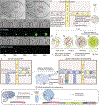
A) (Top) Stochastic (i.e., Poisson) encapsulation of polystyrene microbeads. (Bottom) Inertially ordered encapsulation with similar λ value, 0.98.[183] B) (Top) Inertial ordering of polystyrene microbeads, upstream of droplet formation for ordered encapsulation shown in (A).[183] (Bottom) Schematic of inertial ordering forces within a microchannel, where FW is the wall-induced lift force, FS is the shear-induced lift force, FP is the repulsive force, and YEQ is the equilibrium focusing position. C) Theoretical cell loading density for different probabilities of multi-cell droplet loading events, assuming Poisson distribution. D) Images of cell ordering progression in curved microchannel and subsequent encapsulation.[190] E) Schematic of microfluidic cell encapsulation device with (1) curved microchannel section for focusing cells upstream of (2) droplet formation.[190] (Inset) Schematic of forces present within a curved microchannel, where FD is the Dean force and FL is the net lift force. (A,B) Reproduced with permission.[183] Copyright 2008, Royal Society of Chemistry. C,D) Reproduced with permission.[190] Copyright 2012, Royal Society of Chemistry. Scale bars denote 100 μm for (A) and (B), and 50 μm for (D).
Figure 8.
Figure 8.
A) Confocal image of an encapsulated chondrocyte, with cell membrane stained red and nucleus blue. The PEGDA encapsulant (black) prevents fluorescently labeled dextran (green) from reaching cellular cargo; scale bar denotes 10 μm.[31] B) (Left) Off-center cell positioning within a Dex-TA microgel, which leads to early cell egress. (Right) Centered cell positioning within a Dex-TA microgel for prolonged protection from host immune response and long-term in vitro cell culture.[25] C) (Left) Image of PEGDA single-cell (green) microgels integrated into a fibrin macrogel preventing permeation of fluorescently labeled 70 kDa dextran (red) while permitting permeation of BSA (middle and right); scale bar denotes 25 μm.[31] Confocal images in (A) and (C) plots are reproduced with permission.[31] Copyright 2016, Wiley-VCH. Off-center and centered cell positions in microgel images are reproduced with permission.[25] Copyright 2017, Wiley-VCH.
Figure 9.
Figure 9.
A) Confocal images of MSCs encapsulated within alginate microgels of 0.41 (top) and 1.00 kPa (bottom) stiffness. Blue, nuclei; green, alginate; red, actin; scale bar, 10 μm. ALP expression for cells in the corresponding groups.[46] B) MSCs encapsulated within 5 and 7.5% w/v TG-PEG hydrogels. ALP expression of MSCs in the corresponding groups after culturing 7 days in differentiation medium.[123] Images of single-cell microgels and box plot of ALP expression are reproduced with permission.[46] Copyright 2016, Springer Nature. Images of single-cell microgels and ALP expression percentage plot are reproduced with permission.[123] Copyright 2017, Royal Society of Chemistry.
Figure 10.
Figure 10.
A) (Top) Bright field images of agarose microgels, with the perimeter traced with red dashed line for clarity, lodged into a microfluidic trapping device. Time-lapse bright field and fluorescence confocal microscopy (GFP expression marking pluripotency) of pluripotent mouse embryonic stem cells continuously perfused with 2i media (middle) and N2B27 media (bottom).[70] B) Workflow of agarose-based droplet microfluidic emulsion PCR.[8] C) Schematic of droplet microfluidics-based Single-Cell Copy Number Variation.[8] Images in (A) are reproduced with permission.[46] Copyright 2018, Springer Nature. Schematics from (B) and (C) are reproduced with permission.[8] Copyright 2019, Wiley-VCH.

Similar articles

Cited by

References

    1. Koboldt DC, Steinberg KM, Larson DE, Wilson RK, Mardis ER, Cell 2013, 155, 27. - PMC - PubMed
    1. Kim ES, Ahn EH, Chung E, Kim DH, Mol. Cells 2013, 36, 477. - PMC - PubMed
    1. Mast FD, Ratushny AV, Aitchison JD, J. Cell Biol 2014, 206, 695. - PMC - PubMed
    1. Yuan GC, Cai L, Elowitz M, Enver T, Fan G, Guo G, Irizarry R, Kharchenko P, Kim J, Orkin S, Quackenbush J, Saadatpour A, Schroeder T, Shivdasani R, Tirosh I, Genome Biol. 2017, 18, 84. - PMC - PubMed
    1. Garcia HG, Berrocal A, Kim YJ, Martini G, Zhao J, Curr. Top. Dev. Biol 2020, 137, 1. - PubMed

LinkOut - more resources