. 2022 Nov 3;10(1):77.
doi: 10.1186/s40364-022-00424-x.

Loss of miR-637 promotes cancer cell stemness via WASH/IL-8 pathway and serves as a novel prognostic marker in esophageal squamous cell carcinoma

Affiliations

Loss of miR-637 promotes cancer cell stemness via WASH/IL-8 pathway and serves as a novel prognostic marker in esophageal squamous cell carcinoma

Mengxing Guo et al. Biomark Res. .

Abstract

Background: Esophageal carcinoma is the highly lethal cancer in the world, predominantly in some areas of East Asia. We previously reported that overexpression of cytoskeleton regulator Wiskott-Aldrich syndrome protein and SCAR Homolog (WASH) associates with poor prognosis of patients with esophageal squamous cell carcinoma (ESCC). However, the molecular mechanism and clinical significance involved in WASH overexpression have not been fully elucidated.

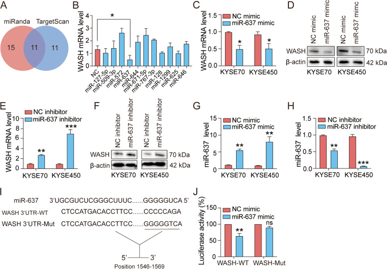
Methods: Bioinformatics analysis and luciferase reporter assay were used to predict and validate miR-637 as a regulator of WASH in ESCC cell lines. qRT-PCR, Western blotting and ELISA assays were performed to examine RNA expression and protein levels, respectively. Next, the biological functions of miR-637 were explored by tumor sphere formation assay in vitro and nude mouse tumor xenograft in vivo. Finally, we evaluated the association of miR-637 levels with clinical features in ESCC patients.

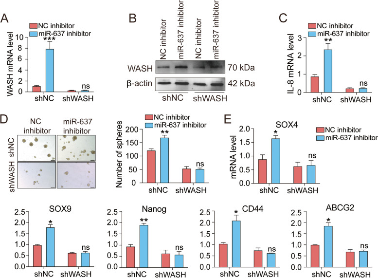
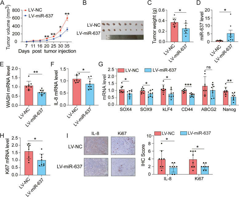
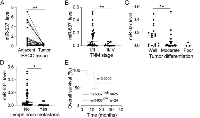
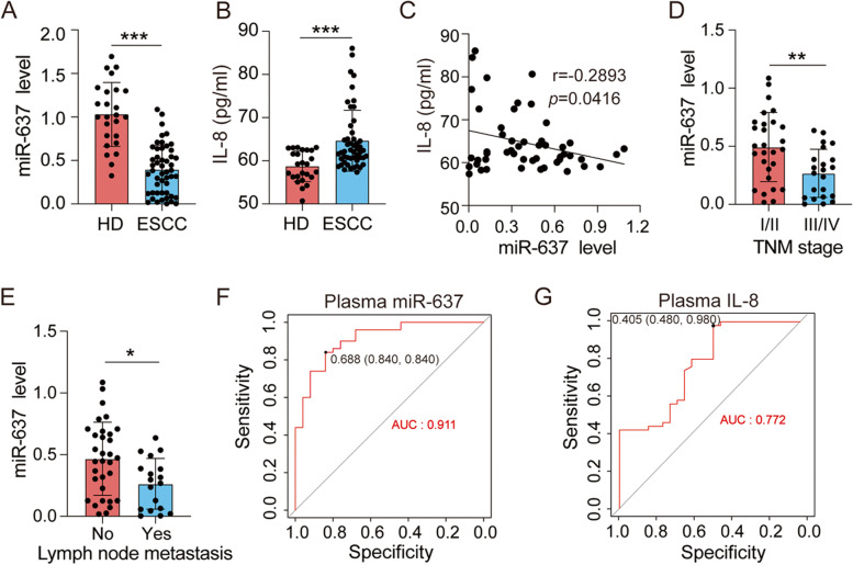
Results: We identified miR-637 as a WASH-targeting miRNA. miR-637 mimic strongly attenuated the downstream IL-8 production and tumor sphere formation in esophageal cancer cells, whereas miR-637 inhibitor displayed an opposite effect. IL-8 could facilitate stem-like properties and partially rescue the phenotypes induced by miR-637 mimic. Furthermore, miR-637 inhibitor dramatically promoted IL-8 expression and cancer stemness properties in a WASH-dependent manner. Ectopic expression of miR-637 also inhibited tumor growth in a mouse model. Clinically, low expression of miR-637 was observed in tumor tissues and the low expression levels of miR-637 were correlated with poor survival of ESCC patients. In particular, plasma miR-637 could be used as a noninvasive biomarker for ESCC patients.

Conclusions: These results implicate the potential application of miR-637 for diagnosis and prognosis of esophageal cancer.

Keywords: Cancer stem cells; Esophageal squamous cell carcinoma; Interleukin-8; WASH; miR-637.


Conflict of interest statement

The authors declare that the research was conducted in the absence of any commercial or financial relationships that could be construed as a potential conflict of interest.

Figures

Fig. 1
Fig. 1
WASH is a direct target of miR-637 in ESCC cells. A The database miRanda and TargetScan were used to predict candidate miRNAs targeting WASH. B Relative expression of WASH was analyzed by qRT-PCR in KYSE70 cells transfected with the miRNA mimics for negative control (NC) or predicted candidates targeting WASH. C, D The down-regulation of WASH expression was determined in both KYSE70 and KYSE450 cells transfected with miR-637 mimic by qRT-PCR (C) and Western blotting (D), respectively. E, F The up-regulation of WASH expression was confirmed in both KYSE70 and KYSE450 cells transfected with miR-637 inhibitor by qRT-PCR (E) and Western blotting (F), respectively. G, H The relative levels of miR-637 were measured by qRT-PCR assays in KYSE70 cells and KYSE450 cells after transfection with mi-637 mimic (G) or miR-637 inhibitor (H), respectively. I Schematic representation of WASH 3′-UTR wild type (WT), and mutant (Mut) with altered residues in the core region of miR-637 binding site. J Luciferase activity in 293 T cells co-transfected with WASH WT-3′-UTR or Mut-3′-UTR constructs and miR-637 or NC mimic, respectively. Data are presented as mean ± standard deviation. ns, not significant; *p < 0.05, **p < 0.01, ***p < 0.001
Fig. 2
Fig. 2
miR-637 suppresses stem-like characteristics in ESCC cells by inhibiting IL-8 expression. A, B qRT-PCR (A) and ELISA (B) assays showed the expression levels of IL-8 in KYSE70 cells and KYSE450 cells after treatment with negative control (NC) mimic or miR-637 mimic. C, D qRT-PCR (C) and ELISA (D) assays showed the expression levels of IL-8 in KYSE70 cells and KYSE450 cells after treatment with NC inhibitor or miR-637 inhibitor. E, F The number of spheres derived from KYSE70 cells and KYSE450 cells after treatment with treated with miR-637 mimic (E) or miR-637 inhibitor (F). G The number of spheres derived from IL-8 overexpressing (OE-IL-8) KYSE450 cells after treatment with miR-637 mimic. Scale bar, 200 μm. Data are presented as mean ± standard deviation. *p < 0.05, **p < 0.01, ***p < 0.001
Fig. 3
Fig. 3
miR-637 inhibits IL-8 expression and cancer stemness in a WASH-dependent manner. KYSE70 cells stably transduced with negative control shRNA (shNC) or WASH shRNA (shWASH) were treated with NC inhibitor or miR-637 inhibitor. A, B qRT-PCR (A) or Western blotting (B) assays showed the failure of miR-637 inhibitor to induce WASH expression in KWSE70 cells with WASH knockdown. C, D qRT-PCR (C) or tumor sphere formation (D) assays showed the failure of miR-637 inhibitor to induce IL-8 expression and sphere formation capacity in KWSE70 cells with WASH knockdown, respectively. Scale bar, 200 μm. E The relative expression of stemness markers were measured by qRT-PCR. Data are presented as mean ± standard deviation. ns, not significant; *p < 0.05, **p < 0.01, ***p < 0.001
Fig. 4
Fig. 4
Ectopic expression of miR-637 reduces the growth of ESCC xenografts in mice. A total of 3 × 106 KYSE450 cells stably transduced with lentivirus (LV) expressing miR-637 or negative control (NC) were implanted subcutaneously in immunodeficient NTG mice (n = 8 per group). A The volumes of transplanted tumors were measured at the indicated time points and the tumor growth curves were plotted. Data are presented as mean ± standard error of mean. B Images of all xenograft tumors excised at day 35 after tumor injection. C All tumor weights were measured. D-H The relative expression of miR-637 (D), WASH (E), IL-8 (F), stemness-related genes (G) and Ki67 (H) were examined by qRT-PCR, respectively. I IHC analysis of IL-8 and Ki67 expression in tumors. Scale bar, 40 μm. Data are presented as mean ± standard deviation. ns, not significant; *p < 0.05, **p < 0.01, ***p < 0.001
Fig. 5
Fig. 5
Low miR-637 expression in tumor tissues is associated with poor pathological features and overall survival of ESCC patients. A The relative expression of miR-637 was examined by qRT-PCR from paired tumor and adjacent normal tissues of 49 ESCC patients. B-D Decreased expression levels of miR-637 in tumor tissues correlate with advanced TNM stage (B), poor differentiation (C), and lymph node metastasis (D), respectively. E Kaplan-Meier survival analysis for overall survival was performed, comparing the relatively low and high miR-637 expression groups of ESCC patients. Data are presented as mean ± standard deviation. *p < 0.05, **p < 0.01
Fig. 6
Fig. 6
Plasma miR-637 is a useful biomarker for the diagnosis and prognosis of ESCC patients. A, B The expression levels of miR-637 and IL-8 in the plasma of healthy donors (HD) and ESCC patients were examined by qRT-PCR (A) and ELISA (B), respectively. C The relationship between miR-637 and IL-8 expression in the plasma of ESCC patients was evaluated via Pearson correlation analysis. D, E Low expression levels of plasma miR-637 correlate with advanced TNM stage (D) and lymph node metastasis (E). F, G The prediction performance of plasma miR-637 (F) or plasma IL-8 (G) in a cohort of ESCC patients were determined by ROC analysis. Data are presented as mean ± standard deviation. ns, not significant; *p < 0.05, **p < 0.01, ***p < 0.001

Similar articles

Cited by

References

    1. Bray F, Ferlay J, Soerjomataram I, Siegel RL, Torre LA, Jemal A. Global cancer statistics 2018: GLOBOCAN estimates of incidence and mortality worldwide for 36 cancers in 185 countries. CA Cancer J Clin. 2018;68(6):394–424. doi: 10.3322/caac.21492. - DOI - PubMed
    1. Arnold M, Soerjomataram I, Ferlay J, Forman D. Global incidence of oesophageal cancer by histological subtype in 2012. Gut. 2015;64(3):381–387. doi: 10.1136/gutjnl-2014-308124. - DOI - PubMed
    1. Thrift AP. Barrett's esophagus and esophageal adenocarcinoma: how common are they really? Dig Dis Sci. 2018;63(8):1988–1996. doi: 10.1007/s10620-018-5068-6. - DOI - PubMed
    1. Dong J, Gu X, El-Serag HB, Thrift AP. Underuse of surgery accounts for racial disparities in esophageal Cancer survival times: a matched cohort study. Clin Gastroenterol Hepatol. 2019;17(4):657–65.e13. doi: 10.1016/j.cgh.2018.07.018. - DOI - PubMed
    1. Rustgi AK, El-Serag HB. Esophageal carcinoma. N Engl J Med. 2014;371(26):2499–2509. doi: 10.1056/NEJMra1314530. - DOI - PubMed

LinkOut - more resources