. 2022 Nov;11(21):e026174.
doi: 10.1161/JAHA.122.026174. Epub 2022 Oct 31.

Myotubularin-Related Protein14 Prevents Neointima Formation and Vascular Smooth Muscle Cell Proliferation by Inhibiting Polo-Like Kinase1

Affiliations

Myotubularin-Related Protein14 Prevents Neointima Formation and Vascular Smooth Muscle Cell Proliferation by Inhibiting Polo-Like Kinase1

Ling-Yao Kong et al. J Am Heart Assoc. 2022 Nov.

Abstract

Background Restenosis is one of the main bottlenecks in restricting the further development of cardiovascular interventional therapy. New signaling molecules involved in the progress have continuously been discovered; however, the specific molecular mechanisms remain unclear. MTMR14 (myotubularin-related protein 14) is a novel phosphoinositide phosphatase that has a variety of biological functions and is involved in diverse biological processes. However, the role of MTMR14 in vascular biology remains unclear. Herein, we addressed the role of MTMR14 in neointima formation and vascular smooth muscle cell (VSMC) proliferation after vessel injury. Methods and Results Vessel injury models were established using SMC-specific conditional MTMR14-knockout and -transgenic mice. Neointima formation was assessed by histopathological methods, and VSMC proliferation and migration were assessed using fluorescence ubiquitination-based cell cycle indicator, transwell, and scratch wound assay. Neointima formation and the expression of MTMR14 was increased after injury. MTMR14 deficiency accelerated neointima formation and promoted VSMC proliferation after injury, whereas MTMR14 overexpression remarkably attenuated this process. Mechanistically, we demonstrated that MTMR14 suppressed the activation of PLK1 (polo-like kinase 1) by interacting with it, which further leads to the inhibition of the activation of MEK/ERK/AKT (mitogen-activated protein kinase kinase/extracellular-signal-regulated kinase/protein kinase B), thereby inhibiting the proliferation of VSMC from the medial to the intima and thus preventing neointima formation. Conclusions MTMR14 prevents neointima formation and VSMC proliferation by inhibiting PLK1. Our findings reveal that MTMR14 serves as an inhibitor of VSMC proliferation and establish a link between MTMR14 and PLK1 in regulating VSMC proliferation. MTMR14 may become a novel potential therapeutic target in the treatment of restenosis.

Keywords: MTMR14; PLK1; VSMC; neointima formation; proliferation.


Figures
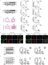
Figure 1
Figure 1. MTMR14 (myotubularin‐related protein 14) is upregulated in proliferating vascular smooth muscle cells (VSMCs) both in vivo and in vitro.
A and B, Representative protein (A) and mRNA (B) levels of MTMR14 in carotid artery of rats 28 days after balloon injury (n=3); (C and D) Representative protein (C) and mRNA (D) levels of MTMR14 in carotid artery of mice 28 days after ligation (n=3); (E) Representative images of hematoxylin–eosin (HE) staining of carotid arteries in rats at 28 days after balloon injury (scale bar, 200 μm; n=6); (F) Representative images of HE staining of carotid arteries in mice at 28 days after ligation (scale bar, 100 μm; n=6); (G) Representative images of immunofluorescence staining for MTMR14 (green), ACTA2 (red) and DAPI (blue) of carotid arteries in rats at 28 days after balloon injury (scale bar, 200 μm); (H) Representative images of immunofluorescence staining for MTMR14 (green), ACTA2 (red), and DAPI (blue) of carotid arteries in mice at 28 days after ligation (scale bar, 100 μm); (I and J) Representative protein (I) and mRNA (J) levels of MTMR14, PCNA, and ACTA2 in primary VSMCs cultured at different platelet‐derived growth factor BB (PDGF‐BB) concentrations (n=3); (K and L) Representative protein (K) and mRNA (L) levels of MTMR14, PCNA, and ACTA2 in primary VSMCs cultured with PDGF‐BB (20 ng/mL) at different time gradients. (*P<0.05 vs sham; **P<0.05 vs 0 ng/mL; ***P<0.05 vs 0 hour; n=3). ACTA2 indicates aortic alpha‐actin; CAL, carotid artery ligation; IF, immunofluorescence; PCNA, proliferating cell nuclear antigen; and PDGF‐BB, platelet‐derived growth factor BB.
Figure 2
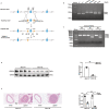
Figure 2. MTMR14 (myotubularin‐related protein 14) deficiency aggravates neointima formation.
A, Schematic workflow for the construction of an SMC‐specific conditional MTMR14‐knockout (Mtmr14Δ) mouse strain; (B) Representative PCR amplification‐mediated genotyping of MTMR14‐loxP/loxP, MTMR14+/+, and MTMR14‐loxP/+ mice; (C) Mouse genotyping was confirmed by PCR; (D) Representative protein level of MTMR14 in carotid arteries of Mtmr14 WT and Mtmr14Δ mice (n=4); (E) Representative hematoxylin–eosin (HE) staining images of carotid arteries in Mtmr14 WT and Mtmr14Δ mice at 28 days after ligation (scale bar, 100 μm; n=6). (*P<0.05 vs sham, **P<0.05 vs Mtmr14 WT). PCR indicates polymerase chain reaction; and SMC, smooth muscle cell.
Figure 3
Figure 3. Overexpression of MTMR14 (myotubularin‐related protein 14) prevents neointima formation.
A, Schematic workflow for the construction of a SMC‐specific conditional MTMR14‐transgenic (Mtmr14 TG) mouse strain; (B) Representative protein level of MTMR14 in carotid arteries of Mtmr14 WT and Mtmr14 TG mice (n=4); (C) Representative hematoxylin–eosin (HE) staining images of carotid arteries in Mtmr14 WT and Mtmr14 TG mice at 28 days after ligation (scale bar, 100 μm; n=6). (*P<0.05 vs sham, **P<0.05 vs Mtmr14 WT).
Figure 4
Figure 4. The effect of MTMR14 (myotubularin‐related protein 14) on proliferation and migration in primary vascular smooth muscle cells (VSMCs) stimulated by platelet‐derived growth factor BB (PDGF‐BB).
A, Representative image of primary VSMCs stably expressing fluorescence ubiquitination‐based cell cycle indicator (FUCCI) (red for G1, yellow for G1/S, green for S/G2‐M) 48 hours after infection of Ad‐shScramble or Ad‐shMTMR14 and stimulated by 20 ng/mL PDGF‐BB; (B and C) Transwell and scratch wound assays of primary VSMCs infected with Ad‐shScramble or Ad‐shMTMR14 after 20 ng/mL PDGF‐BB stimulation for 24 hours; (D) Representative image of primary VSMCs stably expressing FUCCI cell cycle indicators (red for G1, yellow for G1/S, green for S/G2‐M) 48 hours after infection of Ad‐Control or Ad‐MTMR14 and stimulated by 20 ng/mL PDGF‐BB; (E and F) Transwell and scratch wound assays of primary VSMCs infected with Ad‐Control or Ad‐MTMR14 after 20 ng/mL PDGF‐BB stimulation for 24 hours. (scale bar, 100 μm; n=3; *P<0.05 vs Ad‐shScramble, **P<0.05 vs Ad‐Control).
Figure 5
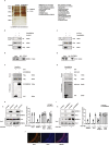
Figure 5. MTMR14 (myotubularin‐related protein 14) can bind to PLK1 (polo‐like kinase 1).
A, Silver‐stained gel of the indicated proteins binding to MTMR14, which were coimmunoprecipitated using an anti‐MTMR14 antibody and identified via mass spectrometry in primary vascular smooth muscle cells (VSMCs). IgG was used as a control; (B and C) 293T cells were transfected with plasmids expressing Flag‐MTMR14 and HA‐PLK1, and immunoprecipitation and Western blot assays was used to detect the binding of MTMR14 and PLK1; (D and E) immunoprecipitation and western blot assays using anti‐MTMR14, and anti‐PLK1 to detect the binding of MTMR14 to PLK1 in primary VSMCs. IgG was used as a control; (F and G) GST pull‐down assays using anti‐GST and anti‐Flag to detect the direct binding of MTMR14 and PLK1. Purified GST was used as a control; (H and I) Immunoprecipitation and Western blot assays using anti‐MTMR14, and anti‐PLK1 to detect the binding of MTMR14 to PLK1 in primary VSMCs stimulated with or without platelet‐derived growth factor BB (PDGF‐BB). IgG was used as a control. J, Representative images of immunofluorescence staining with an anti‐PLK1 antibody in slices from human restenosis artery (scale bar, 100 μm). GST, glutathione S‐transferase; and LC‐MS/MS, liquid chromatography–tandem mass spectrometry.
Figure 6
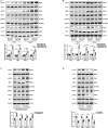
Figure 6. The impact of MTMR14 (myotubularin‐related protein 14) on PLK1 (polo‐like kinase 1) is mediated by inhibiting the phosphorylation of PLK1.
A, Western blot assays were performed in the carotid arteries tissues in Mtmr14 WT or MTMR14‐knockout (Mtmr14Δ) mice at 28 days after ligation to detect the expression of PLK1, p‐PLK1, MEK, p‐MEK, ERK1/2, p‐ERK1/2, AKT, and p‐AKT; (B) Western blot assays were performed in the carotid arteries in Mtmr14 WT or MTMR14‐transgenic (Mtmr14 TG) mice at 28 days after ligation to detect the expression of PLK1, p‐PLK1, MEK, p‐MEK, ERK1/2, p‐ERK1/2, AKT, and p‐AKT; (C) Western blot assays were performed in primary vascular smooth muscle cells (VSMCs) infected with Ad‐shScramble or Ad‐shMTMR14 at 24 hours after platelet‐derived growth factor BB (PDGF‐BB) stimulation to detect the expression of PLK1, p‐PLK1, MEK, p‐MEK, ERK1/2, p‐ERK1/2, AKT, and p‐AKT; (D) Western blot assays were performed in primary VSMCs infected with Ad‐Control or Ad‐MTMR14 at 24 hours after PDGF‐BB stimulation to detect the expression of PLK1, p‐PLK1, MEK, p‐MEK, ERK1/2, p‐ERK1/2, AKT, and p‐AKT. (GAPDH served as the loading control, n=6, *P<0.05 vs sham, **P<0.05 vs Mtmr14 WT # P<0.05 vs Ad‐shScramble, ## P<0.05 vs Ad‐Control). AKT indicates protein kinase B; ERK, extracellular‐signal‐regulated kinase; and MEK, mitogen‐activated protein kinase kinase.
Figure 7
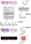
Figure 7. Knockdown PLK1 (polo‐like kinase 1) reversed the neointima formation aggravated by MTMR14 (myotubularin‐related protein 14) knockdown.
A, Representative hematoxylin–eosin (HE) staining images of carotid arteries from Mtmr14 WT or MTMR14‐knockout (Mtmr14Δ) mice local infected with Ad‐shScramble or Ad‐shPLK1 at 28 days after ligation (scale bar, 100 μm; n=6); (B) Western blot assays were performed in primary vascular smooth muscle cells (VSMCs) infected with Ad‐shScramble+Ad‐shScramble, Ad‐shMTMR14+ Ad‐shScramble, Ad‐shScramble+Ad‐PLK1, or Ad‐shMTMR14+ Ad‐shPLK1 at 24 hours after 20 ng/mL platelet‐derived growth factor BB (PDGF‐BB) stimulation to detect the expression of PLK1, p‐PLK1, MEK, p‐MEK, ERK1/2, p‐ERK1/2, AKT, and p‐AKT (GAPDH served as the loading control, n=6); (C) Western blot assays were performed in primary VSMCs infected with Ad‐shScramble+ Ad‐shScramble, Ad‐shMTMR14+ Ad‐shScramble, Ad‐shMTMR14+ Ad‐shPLK1 or Ad‐shMTMR14+ Ad‐shPLK1 after 20 ng/mL PDGF‐BB stimulation for 24 hours to detect the expression of PCNA and ACTA2 (GAPDH served as the loading control, n=6); (D) Transwell assays of primary VSMCs infected with Ad‐shScramble+ Ad‐shScramble, Ad‐shMTMR14+ Ad‐shScramble, Ad‐shMTMR14+ Ad‐shPLK1 or Ad‐shMTMR14+ Ad‐shPLK1 after 20 ng/mL PDGF‐BB stimulation for 24 hours (scale bar, 100 μm; n=3); (E) Representative image of primary VSMCs stably expressing fluorescence ubiquitination‐based cell cycle indicator (FUCCI) (red for G1, yellow for G1/S, green for S/G2‐M) 48 hours after infection of Ad‐shScramble+ Ad‐shScramble, Ad‐shMTMR14+ Ad‐shScramble, Ad‐shMTMR14+ Ad‐shPLK1 or Ad‐shMTMR14+ Ad‐shPLK1 and stimulated by 20 ng/mL PDGF‐BB (scale bar, 100 μm). (*P<0.05 Mtmr14Δ vs Mtmr14 WT, **P<0.05 Ad‐shPLK1 vs Ad‐shScramble, ***P<0.05 Ad‐shMTMR14 vs Ad‐shScramble). ACTA2 indicates aortic alpha‐actin; AKT, protein kinase B; ERK, extracellular‐signal‐regulated kinase; MEK, mitogen‐activated protein kinase kinase; and PCNA, proliferating cell nuclear antigen.
Figure 8
Figure 8. MTMR14 (myotubularin‐related protein 14) prevents neointima formation and vascular smooth muscle cell proliferation by inhibiting PLK1/MEK/ERK/AKT axis.
PLK1/MEK/ERK/AKT axis was activated in vascular smooth muscle cells (VSMCs) after injury promoting VSMC proliferation and neointima formation. MTMR14 inhibited the activation of PLK1 (polo‐like kinase 1) by interacting with PLK1, which further inhibited the activation of the MEK/ERK/AKT axis, thereby inhibiting VSMC proliferation and neointima formation. ACTA2 indicates aortic alpha‐actin; AKT, protein kinase B; CAL, carotid artery ligation; ERK, extracellular‐signal‐regulated kinase; FUCCI, fluorescence ubiquitination‐based cell cycle indicator; HE, hematoxylin–eosin; MEK, mitogen‐activated protein kinase kinase; PCNA, proliferating cell nuclear antigen; and PDGF‐BB, platelet‐derived growth factor BB.

Similar articles

Cited by

References

    1. DALYs GBD and Collaborators H . Global, regional, and national disability‐adjusted life‐years (DALYs) for 333 diseases and injuries and healthy life expectancy (HALE) for 195 countries and territories, 1990–2016: a systematic analysis for the Global Burden of Disease Study 2016. Lancet. 2017;390:1260–1344. doi: 10.1016/S0140-6736(17)32130-X - DOI - PMC - PubMed
    1. Collaborators GBDCoD . Global, regional, and national age‐sex specific mortality for 264 causes of death, 1980–2016: a systematic analysis for the Global Burden of Disease Study 2016. Lancet. 2017;390:1151–1210. doi: 10.1016/S0140-6736(17)32152-9 - DOI - PMC - PubMed
    1. Durham AL, Speer MY, Scatena M, Giachelli CM, Shanahan CM. Role of smooth muscle cells in vascular calcification: implications in atherosclerosis and arterial stiffness. Cardiovasc Res. 2018;114:590–600. doi: 10.1093/cvr/cvy010 - DOI - PMC - PubMed
    1. Bennett MR, Sinha S, Owens GK. Vascular smooth muscle cells in atherosclerosis. Circ Res. 2016;118:692–702. doi: 10.1161/CIRCRESAHA.115.306361 - DOI - PMC - PubMed
    1. Grootaert MOJ, Moulis M, Roth L, Martinet W, Vindis C, Bennett MR, De Meyer GRY. Vascular smooth muscle cell death, autophagy and senescence in atherosclerosis. Cardiovasc Res. 2018;114:622–634. doi: 10.1093/cvr/cvy007 - DOI - PubMed

Publication types

MeSH terms

Substances

LinkOut - more resources