Epigenetics of neural differentiation: Spotlight on enhancers
- PMID: 36313573
- PMCID: PMC9606577
- DOI: 10.3389/fcell.2022.1001701
Epigenetics of neural differentiation: Spotlight on enhancers
Abstract
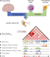
Neural induction, both in vivo and in vitro, includes cellular and molecular changes that result in phenotypic specialization related to specific transcriptional patterns. These changes are achieved through the implementation of complex gene regulatory networks. Furthermore, these regulatory networks are influenced by epigenetic mechanisms that drive cell heterogeneity and cell-type specificity, in a controlled and complex manner. Epigenetic marks, such as DNA methylation and histone residue modifications, are highly dynamic and stage-specific during neurogenesis. Genome-wide assessment of these modifications has allowed the identification of distinct non-coding regulatory regions involved in neural cell differentiation, maturation, and plasticity. Enhancers are short DNA regulatory regions that bind transcription factors (TFs) and interact with gene promoters to increase transcriptional activity. They are of special interest in neuroscience because they are enriched in neurons and underlie the cell-type-specificity and dynamic gene expression profiles. Classification of the full epigenomic landscape of neural subtypes is important to better understand gene regulation in brain health and during diseases. Advances in novel next-generation high-throughput sequencing technologies, genome editing, Genome-wide association studies (GWAS), stem cell differentiation, and brain organoids are allowing researchers to study brain development and neurodegenerative diseases with an unprecedented resolution. Herein, we describe important epigenetic mechanisms related to neurogenesis in mammals. We focus on the potential roles of neural enhancers in neurogenesis, cell-fate commitment, and neuronal plasticity. We review recent findings on epigenetic regulatory mechanisms involved in neurogenesis and discuss how sequence variations within enhancers may be associated with genetic risk for neurological and psychiatric disorders.
Keywords: cell-type specific; enhancers; epigenetics; neural induction; neurogenesis; transcription regulation.
Copyright © 2022 Giacoman-Lozano, Meléndez-Ramírez, Martinez-Ledesma, Cuevas-Diaz Duran and Velasco.
Conflict of interest statement
The authors declare that the research was conducted in the absence of any commercial or financial relationships that could be construed as a potential conflict of interest.
Figures


Similar articles
-
Dynamics and function of distal regulatory elements during neurogenesis and neuroplasticity.Genome Res. 2015 Sep;25(9):1309-24. doi: 10.1101/gr.190926.115. Epub 2015 Jul 13. Genome Res. 2015. PMID: 26170447 Free PMC article.
-
Genetics and Epigenetics in Adult Neurogenesis.Cold Spring Harb Perspect Biol. 2016 Jun 1;8(6):a018911. doi: 10.1101/cshperspect.a018911. Cold Spring Harb Perspect Biol. 2016. PMID: 27143699 Free PMC article. Review.
-
Epigenetics, hippocampal neurogenesis, and neuropsychiatric disorders: unraveling the genome to understand the mind.Neurobiol Dis. 2010 Jul;39(1):73-84. doi: 10.1016/j.nbd.2010.01.008. Epub 2010 Jan 28. Neurobiol Dis. 2010. PMID: 20114075 Free PMC article. Review.
-
Bridging between Mouse and Human Enhancer-Promoter Long-Range Interactions in Neural Stem Cells, to Understand Enhancer Function in Neurodevelopmental Disease.Int J Mol Sci. 2022 Jul 19;23(14):7964. doi: 10.3390/ijms23147964. Int J Mol Sci. 2022. PMID: 35887306 Free PMC article.
-
Opening up the blackbox: an interpretable deep neural network-based classifier for cell-type specific enhancer predictions.BMC Syst Biol. 2016 Aug 1;10 Suppl 2(Suppl 2):54. doi: 10.1186/s12918-016-0302-3. BMC Syst Biol. 2016. PMID: 27490187 Free PMC article.
Cited by
-
Hybrid Epigenomes Reveal Extensive Local Genetic Changes to Chromatin Accessibility Contribute to Divergence in Embryonic Gene Expression Between Species.Mol Biol Evol. 2023 Nov 3;40(11):msad222. doi: 10.1093/molbev/msad222. Mol Biol Evol. 2023. PMID: 37823438 Free PMC article.
-
Distinct disease mutations in DNMT3A result in a spectrum of behavioral, epigenetic, and transcriptional deficits.bioRxiv [Preprint]. 2023 Feb 27:2023.02.27.530041. doi: 10.1101/2023.02.27.530041. bioRxiv. 2023. Update in: Cell Rep. 2023 Nov 28;42(11):113411. doi: 10.1016/j.celrep.2023.113411. PMID: 36909558 Free PMC article. Updated. Preprint.
-
Enhancing Our Understanding of Cell Types, Differentiation, and Disease Through Enhancers: An Interview with César Daniel Meléndez-Ramírez, MS.Yale J Biol Med. 2023 Dec 29;96(4):569-572. eCollection 2023 Dec. Yale J Biol Med. 2023. PMID: 38161574 Free PMC article. No abstract available.
-
An Overview of the Epigenetic Modifications in the Brain under Normal and Pathological Conditions.Int J Mol Sci. 2024 Mar 30;25(7):3881. doi: 10.3390/ijms25073881. Int J Mol Sci. 2024. PMID: 38612690 Free PMC article. Review.
-
Distinct disease mutations in DNMT3A result in a spectrum of behavioral, epigenetic, and transcriptional deficits.Cell Rep. 2023 Nov 28;42(11):113411. doi: 10.1016/j.celrep.2023.113411. Epub 2023 Nov 11. Cell Rep. 2023. PMID: 37952155 Free PMC article.
References
Publication types
LinkOut - more resources
Full Text Sources

