Review
. 2022 Oct 21;11(20):3317.
doi: 10.3390/cells11203317.

Therapeutic Targeting of NF-κB in Acute Lung Injury: A Double-Edged Sword

Affiliations
Review

Therapeutic Targeting of NF-κB in Acute Lung Injury: A Double-Edged Sword

Michelle Warren Millar et al. Cells. .

Abstract

Acute lung injury/acute respiratory distress syndrome (ALI/ARDS) is a devastating disease that can be caused by a variety of conditions including pneumonia, sepsis, trauma, and most recently, COVID-19. Although our understanding of the mechanisms of ALI/ARDS pathogenesis and resolution has considerably increased in recent years, the mortality rate remains unacceptably high (~40%), primarily due to the lack of effective therapies for ALI/ARDS. Dysregulated inflammation, as characterized by massive infiltration of polymorphonuclear leukocytes (PMNs) into the airspace and the associated damage of the capillary-alveolar barrier leading to pulmonary edema and hypoxemia, is a major hallmark of ALI/ARDS. Endothelial cells (ECs), the inner lining of blood vessels, are important cellular orchestrators of PMN infiltration in the lung. Nuclear factor-kappa B (NF-κB) plays an essential role in rendering the endothelium permissive for PMN adhesion and transmigration to reach the inflammatory site. Thus, targeting NF-κB in the endothelium provides an attractive approach to mitigate PMN-mediated vascular injury, not only in ALI/ARDS, but in other inflammatory diseases as well in which EC dysfunction is a major pathogenic mechanism. This review discusses the role and regulation of NF-κB in the context of EC inflammation and evaluates the potential and problems of targeting it as a therapy for ALI/ARDS.

Keywords: endothelial cells; lung inflammatory injury; signal transduction; transcription factors.


Conflict of interest statement

The authors do not claim any conflicts of interest.

Figures

Figure 1
Figure 1
The capillary-alveolar unit in healthy and injured lung. (A) In the healthy lung, the intact capillary-alveolar unit (endothelium and epithelium) prevents the influx of immune cells and fluid into the interstitium and alveoli. (B) In the injured lung, disruption of the endothelial and epithelial barriers causes pulmonary edema, inflammatory cell recruitment (primarily polymorphonuclear leukocytes (PMNs)), and hypoxemia.
Figure 2
Figure 2
PMN adhesion and transendothelial migration (TEM). (A) Resting PMNs express beta-2 (β2) integrins and selectin ligands, but unstimulated endothelial cells (ECs) do not express selectins and are, thus, unable to bind circulating PMNs. (B) Microbial infections and proinflammatory mediators stimulate the endothelium, and activated ECs express cell surface adhesion molecules (P-selectin, E-selectin, and intercellular adhesion molecule-1 (ICAM-1)), which interact with their counter receptors on PMNs. Primary PMN capture and secondary PMN–PMN tethering are facilitated by interactions between selectins and selectin ligands. As captured PMNs slow in circulation and roll along the endothelium, β2 integrins are activated. (C) Activated β2 integrins interact with EC ICAM-1, which clusters to promote firm EC-PMN adhesion. β2-integrin/ICAM-1 interaction facilitates PMN spreading and crawling, and eventually, PMN transendothelial migration.
Figure 3
Figure 3
NF-κB activators and cell responses. Nuclear factor-kappa B (NF-κB) is activated by proinflammatory stimuli including cytokines (tumor necrosis factor alpha (TNFα), interleukin-1 beta (IL-1β)), microbial infection (bacteria/lipopolysaccharide (LPS), viruses), receptor ligands (thrombin, CD40 ligand (CD40L), B-cell-activating factor (BAFF)), and stress (endoplasmic reticulum (ER) stress, reactive oxygen species (ROS)). Stimulation liberates the NF-κB dimer secondary to degradation of IκBα (inhibitor of κB) in the cytosol and promotes its nuclear translocation and DNA binding to activate transcription of genes involved in numerous cell responses.
Figure 4
Figure 4
Dysregulated NF-κB signaling is implicated in the progression of numerous disease states.
Figure 5
Figure 5
Structure of NF-κB family members. The number of amino acids in each protein is listed on the right. (A) The five members of the NF-κB family contain a Rel homology domain (RHD) at their N-terminus. The RHD is comprised of a DNA binding domain, a dimerization/IκB binding domain, and a nuclear localization sequence (NLS). At their C-terminus, the catalytically active RelA, RelB, and c-Rel proteins contain transactivation domains (TADs), whereas p105 and p100 contain a glycine-rich region (GRR) and ankyrin repeats (ovals). The p105 and p100 proteins are catalytically inactive due to the presence of their ankyrin repeats, and the GRR facilitates the cotranslational processing of p105 to p50 and the post-translational processing of p100 to p52. (B) The ankyrin repeats (ovals) in IκB proteins interact with the RHD of NF-κB family members and mask its NLS, and serine phosphorylation at the indicated sites promotes their proteasomal degradation. BCL-3 also contains a TAD at its C-terminus, which confers transcriptional activity to p50/BCL-3 and p52/BCL-3 complexes. (C) The catalytically active IKKα and IKKβ proteins contain kinase and helix–loop–helix (HLH) domains, and their activity is stimulated by phosphorylation of the kinase domain at the indicated serines. The regulatory protein, IKKγ (or NEMO), contains a zinc finger (Z) domain, two coiled-coil domains, and two a-helical (a) domains, which mediate its association with the NEMO binding domain (NBD) at the C-terminus of IKKα and IKKβ. All three members also contain a leucine zipper (LZ) motif required for protein–protein interaction. (D) NF-κB-inducing kinase (NIK) activates IKKα, which phosphorylates the p100 C-terminus to stimulate the post-translational processing of p100 to p52. The p100 protein is cleaved at amino acid 447, and the C-terminus is degraded by the 26S proteasome. The GRR serves as a signal to prevent the proteasome from degrading the N-terminal portion of the protein.
Figure 6
Figure 6
Canonical and noncanonical NF-κB pathways. (Left) The canonical pathway is activated by proinflammatory stimuli including TNFα, thrombin, IL-1β, and LPS, among others. Interaction of extracellular stimuli with cell surface receptors results in activation of the IκB kinase (IKK) complex, including the catalytic subunits IKKα and IKKβ. These subunits phosphorylate IκBα, which leads to its ubiquitination and degradation, exposing the NLS on the NF-κB dimer. The NF-κB dimer, predominantly a heterodimer of p50-RelA/p65, translocates to the nucleus and binds DNA to stimulate the transcription of innate immunity, inflammatory, and cell survival genes. (Right) The noncanonical pathway is activated by BAFF, lymphotoxin-β (LTβ), CD40L, and LPS, which signal through NF-κB-inducing kinase (NIK) to activate the IKKα homodimer. IKKα phosphorylates the NF-κB protein p100 to stimulate its processing into p52. This processing exposes the NLS on p52, allowing p52/RelB heterodimers to translocate to the nucleus and activate gene transcription. The noncanonical pathway primarily regulates expression of genes involved in B cell maturation and lymphoid organogenesis. (P denotes protein phosphorylation).
Figure 7
Figure 7
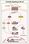
Thrombin-activated NF-κB pathway in endothelial cells. Thrombin activates its receptor, the G-protein-coupled receptor (GPCR) protease-activated receptor (PAR-1), by cleaving the extracellular domain to stimulate signaling. The Gβγ subunit activates phosphatidylinositol-3 kinase (PI3K)/Akt, which phosphorylates the IKK complex to activate canonical NF-κB signaling and induce RelA/p65 homodimer translocation to the nucleus. Gαq phosphorylates protein kinase C delta (PKCδ), which activates NF-κB by dual mechanisms. It induces IKK complex phosphorylation to release RelA/p65 for its translocation and DNA binding in the nucleus. It also engages p38 and c-Src/spleen tyrosine kinase (Syk) to phosphorylate RelA/p65 and enhance its transcriptional activity. Whereas p38 causes serine phosphorylation (red), c-Src and Syk cause tyrosine phosphorylation (purple).
Figure 8
Figure 8
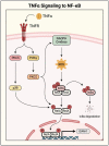
NF-κB activation by TNFα in endothelial cells. TNFα binding to its receptor, TNF receptor (TNFR), activates NF-κB by parallel pathways. It signals through PI3Kγ to activate PKCζ, which triggers NADPH oxidase to produce ROS. ROS then causes phosphorylation and degradation of IκBα and liberates RelA/p65 for its nuclear translocation and DNA binding. TNFα also initiates RelA/p65 serine phosphorylation through PI3Kγ/PKCζ and PKCδ/p38 pathways, amplifying its transcriptional activity.
Figure 9
Figure 9
Small GTPase/cytoskeletal regulation of thrombin-induced NF-κB activation. Thrombin activation of PAR-1 stimulates RhoA/Rho-associated kinase (ROCK) signaling, nonmuscle myosin light-chain kinase (nmMLCK) activity, and autophagic flux. RhoA/ROCK activates LIM kinase 1 (LIMK1), which acts to phosphorylate IKKβ, causing release of RelA/p65 from IκBα. In parallel, LIMK1 causes phosphorylation and inactivation of cofilin-1 to induce actin cytoskeleton reorganization. Thrombin also activates nmMLCK, which acts in concert with LIMK1/cofilin-1 to promote actin–myosin interaction. Generation of actin–myosin stress fibers by this mechanism facilitates the nuclear translocation of released RelA/p65. Induction of autophagy via Beclin-1 and ATG7 also contributes to cofilin-1 phosphorylation and stress fiber formation. Cofilin-1 phosphorylation is tightly regulated by engagement of slingshot-1Long (SSH-1L), a cofilin-1 phosphatase.
Figure 10
Figure 10
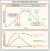
Roles of NF-κB signaling in evolution and resolution of inflammation. (A) In response to infection and injury, NF-κB activation initiates transcription of proinflammatory, proadhesive, and procoagulant genes. As resolution begins, changes/decline in NF-κB signaling promote tissue repair by inducing anti-inflammatory, antiadhesive, anticoagulant, and prosurvival mechanisms. (B) Dampening inflammation: an effective strategy to treat inflammatory conditions. Healthy inflammation (green) is self-limiting and resolves after infection is eliminated and tissue is repaired. However, in many disease conditions (red) inflammation becomes exuberant and persistent, and exerts detrimental effects on host tissues. Abolishing NF-κB/inflammation (orange) is counterproductive as it would impair the host defense response and tissue repair. Developing effective therapeutics to treat inflammatory conditions relies on selectively targeting detrimental inflammation while leaving healthy inflammation intact.
Figure 11
Figure 11
Restraining NF-κB signaling: A promising therapeutic strategy against inflammatory diseases. (A) Signaling to NF-κB is augmented in the setting of inflammatory diseases and may be regulated via multiple pathways. (B) Direct blockade of the receptor or NF-κB may abolish inflammation, compromising the host defense and tissue repair mechanisms. (C) Identification of signaling networks in control of aberrant NF-κB activation and selective targeting of key signaling molecules (alone or in combination) is key to eliminate the detrimental inflammation while maintaining the host defense, tissue repair, and homeostasis functions.

Similar articles

Cited by

References

    1. Matthay M.A., Zemans R.L., Zimmerman G.A., Arabi Y.M., Beitler J.R., Mercat A., Herridge M., Randolph A.G., Calfee C.S. Acute respiratory distress syndrome. Nat. Rev. Dis. Prim. 2019;5:18. doi: 10.1038/s41572-019-0069-0. - DOI - PMC - PubMed
    1. Perl M., Lomas-Neira J., Venet F., Chung C.S., Ayala A. Pathogenesis of indirect (secondary) acute lung injury. Expert. Rev. Respir. Med. 2011;5:115–126. doi: 10.1586/ers.10.92. - DOI - PMC - PubMed
    1. Jagrosse M.L., Dean D.A., Rahman A., Nilsson B.L. RNAi therapeutic strategies for acute respiratory distress syndrome. Transl. Res. 2019;214:30–49. doi: 10.1016/j.trsl.2019.07.011. - DOI - PMC - PubMed
    1. Tzotzos S.J., Fischer B., Fischer H., Zeitlinger M. Incidence of ARDS and outcomes in hospitalized patients with COVID-19: A global literature survey. Crit. Care. 2020;24:516. doi: 10.1186/s13054-020-03240-7. - DOI - PMC - PubMed
    1. Hasan A., Susanto H., Kasim M.F., Nuraini N., Lestari B., Triany D., Widyastuti W. Superspreading in early transmissions of COVID-19 in Indonesia. Sci. Rep. 2020;10:22386. doi: 10.1038/s41598-020-79352-5. - DOI - PMC - PubMed

Publication types