Review
. 2022 Oct 19;9(10):576.
doi: 10.3390/bioengineering9100576.

Broadening the Horizons of RNA Delivery Strategies in Cancer Therapy

Affiliations
Review

Broadening the Horizons of RNA Delivery Strategies in Cancer Therapy

Shuaiying Wu et al. Bioengineering (Basel). .

Abstract

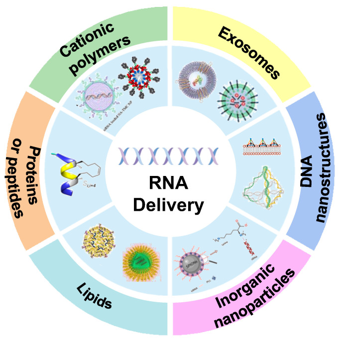
RNA-based therapy is a promising and innovative strategy for cancer treatment. However, poor stability, immunogenicity, low cellular uptake rate, and difficulty in endosomal escape are considered the major obstacles in the cancer therapy process, severely limiting the development of clinical translation and application. For efficient and safe transport of RNA into cancer cells, it usually needs to be packaged in appropriate carriers so that it can be taken up by the target cells and then be released to the specific location to perform its function. In this review, we will focus on up-to-date insights of the RNA-based delivery carrier and comprehensively describe its application in cancer therapy. We briefly discuss delivery obstacles in RNA-mediated cancer therapy and summarize the advantages and disadvantages of different carriers (cationic polymers, inorganic nanoparticles, lipids, etc.). In addition, we further summarize and discuss the current RNA therapeutic strategies approved for clinical use. A comprehensive overview of various carriers and emerging delivery strategies for RNA delivery, as well as the current status of clinical applications and practice of RNA medicines are classified and integrated to inspire fresh ideas and breakthroughs.

Keywords: RNA delivery; cancer therapy; clinical practice; delivery carrier.


Conflict of interest statement

The authors declare no conflict of interest.

Figures

Figure 1
Figure 1
Different RNA delivery carriers in cancer therapy. These delivery vehicles include cationic polymers, exosomes, DNA nanostructures, inorganic nanoparticles, lipids, proteins or peptides, etc.
Figure 2
Figure 2
(A) The formation process of PEIF/siRNA complex and its application in tumor cells. Fluorinated PEIF was synthesized by forming an amide bond between PEI and heptafluorobutyric anhydride. The complex system formed by the combination of PEIF and nucleic acid is stable and easy to be taken up by cells, with high endosomal escape efficiency and gene silencing ability [66]. Copyright 2020, Springer Nature. (B) Schematic illustration shows the PAPD-siRNA and its mode of action in tumor cells. When PAPD nanoparticles are extravasated into tumor tissue, the hypoxic microenvironment causes bio-reducing cleavage of azo adaptor in the activated PAPD complex, resulting in shedding of PEG layer and exposure of the positive charged PEI previously shielded by PEG. This leads to increased uptake of nanoparticles by tumor cells [67]. Copyright 2020, Elsevier. (C) Production and therapeutic effect of PEI/siRNA nanoparticles modified by ECVs. ECV-modified PEI/siRNA complex targeting survivin increased inhibition of PC3 prostate cancer cells Tumor growth curves showed that tumor growth was significantly inhibited by approximately 45% in the treated group [68]. Copyright 2020, Elsevier. (D) TMC synthesis steps and schematic diagram of siRNA loaded HA-TMC NP [26]. Copyright 2020, Elsevier. (E) The fabrication process of siMDR1/DOX co-delivery nanosystems. The nanoplatform consists of three components: the redox-responsive and negatively charged CM-DOX “core”, the pH-responsive dissociated and positively charged “shell” oligoethylene imide /siMDR1, and the surface-modified AS1411 aptam-conjugated hyaluronic acid (AHA) and GALA peptide-conjugated hyaluronic acid (GHA) [69]. Copyright 2022, Elsevier. * p < 0.05, ** p < 0.01.
Figure 3
Figure 3
(A) The diagram describes the NKExos production process diagram. After 72 h of culture, NK92MI cells were removed by differential centrifugation. Finally, NKExos was concentrated and purified [92]. Copyright: © 2021 by the authors. (B) Preparation flow chart of engineered exosome nanocapsules based on 5-FU and Mir-21i. Her2 fuses with LAMP2 to form HCT-116 5FR cells. Her2-lamp2 fusion protein promotes targeted cellular uptake through EGFR receptor-mediated endocytosis in HCT-116 cells. 5-FU and Exo were mixed by electroporation, and 5-FU and Mir-21i were packaged into engineered exosomes and incubated together to form a co-delivery system (THLG-EXO/5-FU/Mir-21I) [93]. Copyright 2020, Springer Nature.
Figure 4
Figure 4
Schematic diagram of the nanobox-siR synthesis (A) and structural transition of its pH-responsive switch (B). The tetrahedral nanobox exoskeleton and siRNA encapsulation and protection were formed simultaneously by one-pot annealing (95 °C for 10 min and 4 °C for 20 min). At pH = 5, the nanobox nucleic acid sequence is transformed into a four-stranded I-motif structure, which subsequently leads to structural disintegration of the spatial tetrahedron and release of the target siRNA [96]. Copyright 2022, Wiley-VCH. (C) Schematic diagram of tFNAS-Cas13a synthesis process and its action on cancer cells. These cells were surface engineered using amphipathic tFNA with three cholesterol-modified vertices and one biotin-modified vertex. T7 polymerase recognizes dsDNA in the T7 promoter region and simultaneously generates a large number of copies of single-stranded RNA. Cas13a recognizes target RNA to activate CRISPR/Cas13a trans cleavage capability, and ssRNA with a fluorophore and quench groups is cleaved by this RNA cleavage capability [97]. Copyright 2022, Elsevier.
Figure 5
Figure 5
(A) Schematic diagram of CaP nanoparticles with different chemical and morphological characteristics and a simplified diagram for the delivery of survivin and cyclin B1-specific siRNA. (B) Through this procedure, acicular hydroxyapatite (HA-N), spherical hydroxyapatite (HA-S) and calcium-deficient hydroxyapatite (CDHA) with average particle sizes of 15, 38, and 20 nm were obtained. The aspect ratio of HA-N and CDHA is about 6. CaP-Arg-siRNAs effectively down-regulated the expression of survivin and cyclin B1 genes to significantly induce cell apoptosis [25]. Copyright 2020, Elsevier. (C) Schematic diagram of gene carrier system of superparamagnetic iron oxide nanoparticles (SPIONs). The consists of SPION coated with the biocompatible protein sericin (Ser) and modified with the cationic polymer polylysine (PLL) to bind negatively charged siRNA [102]. Copyright 2021, Elsevier. (D) Structure diagram of caffeic acid-magnetic calcium phosphate (CAFA-MCAP) nanoparticles. The nucleus consists of superparamagnetic iron oxide nanoparticles (SPION) coated with caffeic acid and stabilized by calcium phosphate, siRNA, and PEG-polyanionic block copolymer layers [103]. Copyright 2020, Elsevier.
Figure 6
Figure 6
(A) Schematic diagram of the DOP-DEDA structure and pH response of DOP-DEDA LNPs, and TEM image (B) of DOP-DEDA LNP. DOP-DEDA has a negatively charged phosphate and two amino groups linked by an ethylene bridge. Produces an almost neutral charge at pH 7.4. The total positive charge at pH = 6.0 and the total negative charge at pH = 8.0 [106]. Copyright 2020, Elsevier. (C) LNPs nanostructure and CL4H6 lipid chemical structure. LNP is composed of CL4H6 lipids, CHOL and PEG-lipids [107]. Copyright 2020, Elsevier. (D) Schematic representation of dendrimer/DOPE/cholesterol/PBD-lipid/mRNA nanoparticles and mRNA delivery process. The combination of PDB lipids and DLNP enhances mRNA production in cancer cells, illuminating tumors by PH-responsive NIR imaging [108]. Copyright 2020, Elsevier.
Figure 7
Figure 7
(A) Peptide synthesis and gene silencing process (B). Fluorescent pin peptides were synthesized by closed-loop metathesis (JMV6337) to encapsulate siRNA and induce gene silencing using short helical pin peptides [110]. Copyright 2020, MDPI. (C) Schematic diagram of APR nanoparticle composition and silencing targeted gene expression. APR nanoparticles are composed of ErbB3 aptamer, protamine and siRNA. After entering cells by ErbB3 aptamer recognition, siRNA silenced the survivin gene and induced apoptosis of ErBB3-positive cells [111]. Copyright 2020, Elsevier. (D) Schematic diagram of EV delivery strategy of siRNA and predicted therapeutic effect on CRPC cells [113]. Copyright 2022, Informa UK Limited.

Similar articles

Cited by

  • Bioengineered nanotechnology for nucleic acid delivery.
    Zhang Y, Luo J, Gui X, Zheng Y, Schaar E, Liu G, Shi J. Zhang Y, et al. J Control Release. 2023 Dec;364:124-141. doi: 10.1016/j.jconrel.2023.10.034. Epub 2023 Oct 27. J Control Release. 2023. PMID: 37879440 Free PMC article. Review.
  • RNA Vaccines: Yeast as a Novel Antigen Vehicle.
    Silva AJD, de Sousa MMG, de Macêdo LS, de França Neto PL, de Moura IA, Espinoza BCF, Invenção MDCV, de Pinho SS, da Gama MATM, de Freitas AC. Silva AJD, et al. Vaccines (Basel). 2023 Aug 7;11(8):1334. doi: 10.3390/vaccines11081334. Vaccines (Basel). 2023. PMID: 37631902 Free PMC article. Review.

References

    1. Zhou J., Rao L., Yu G., Cook T.R., Chen X., Huang F. Supramolecular cancer nanotheranostics. Chem. Soc. Rev. 2021;50:2839–2891. doi: 10.1039/D0CS00011F. - DOI - PubMed
    1. Cheng Z., Li M., Dey R., Chen Y. Nanomaterials for cancer therapy: Current progress and perspectives. J. Hematol. Oncol. 2021;14:85. doi: 10.1186/s13045-021-01096-0. - DOI - PMC - PubMed
    1. Anguela X.M., High K.A. Entering the modern era of gene therapy. Annu. Rev. Med. 2019;70:273–288. doi: 10.1146/annurev-med-012017-043332. - DOI - PubMed
    1. Jeon Y.H., Jung Y.T. Production of a replicating retroviral vector expressing reovirus fast protein for cancer gene therapy. J. Virol. Methods. 2022;299:114332. doi: 10.1016/j.jviromet.2021.114332. - DOI - PubMed
    1. Duan L., Xu L., Xu X., Qin Z., Zhou X., Xiao Y., Liang Y., Xia J. Exosome-mediated delivery of gene vectors for gene therapy. Nanoscale. 2021;13:1387–1397. doi: 10.1039/D0NR07622H. - DOI - PubMed

Grants and funding

China Postdoctoral Science Foundation Funded Project (2021M702743), and the National Natural Science Foundation of China (NSFC, Nos. 81925019, 32101113).

LinkOut - more resources