. 2022 Oct 25;41(4):111545.
doi: 10.1016/j.celrep.2022.111545.

Isoform-specific inhibition of FGFR signaling achieved by a de-novo-designed mini-protein

Affiliations

Isoform-specific inhibition of FGFR signaling achieved by a de-novo-designed mini-protein

Joon Sung Park et al. Cell Rep. .

Abstract

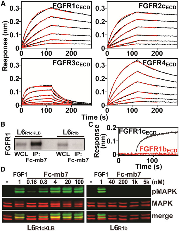
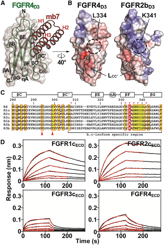
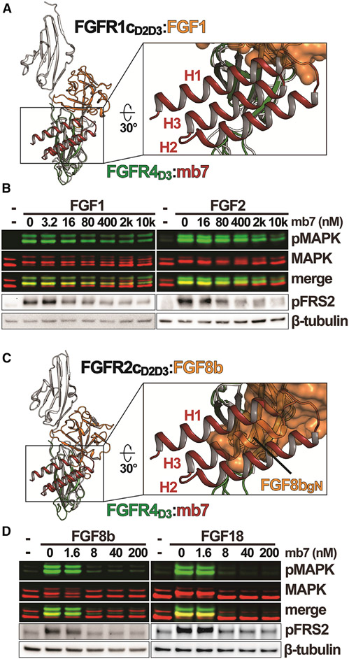
Cellular signaling by fibroblast growth factor receptors (FGFRs) is a highly regulated process mediated by specific interactions between distinct subsets of fibroblast growth factor (FGF) ligands and two FGFR isoforms generated by alternative splicing: an epithelial b- and mesenchymal c-isoforms. Here, we investigate the properties of a mini-protein, mb7, developed by an in silico design strategy to bind to the ligand-binding region of FGFR2. We describe structural, biophysical, and cellular analyses demonstrating that mb7 binds with high affinity to the c-isoforms of FGFR, resulting in inhibition of cellular signaling induced by a subset of FGFs that preferentially activate c-isoforms of FGFR. Notably, as mb7 blocks interaction between FGFR with Klotho proteins, it functions as an antagonist of the metabolic hormones FGF19 and FGF21, providing mechanistic insights and strategies for the development of therapeutics for diseases driven by aberrantly activated FGFRs.

Keywords: CP: Cell biology; cell signaling; de novo protein design; fibroblast growth factors; protein structure; receptor tyrosine kinase.


Conflict of interest statement

Declaration of interests The authors declare no competing interests.

Figures

Figure 1.
Figure 1.. mb7 can potently bind to c-isoforms, but not b-isoforms, of FGFRs
(A) Representative sensorgrams from BLI measurements for the interactions between mb7 and FGFR c-isoforms. Anti-human Fc capture biosensors immobilized with Fc-mb7 were dipped into solutions containing a series of concentrations of FGFR1cECD, FGFR2cECD, FGFR3cECD, or FGFR4ECD. Sensorgrams (black lines) were fitted with a 1:1 binding model (red lines) to calculate binding kinetic parameters. (B) Equal amounts of whole-cell lysates (WCLs) of L6R1cKLB or L6R1b with matching levels of FGFR expressions were incubated with Fc-mb7 immobilized on protein A beads and the bound proteins were immunoblotted using an anti-FGFR1 antibody. (C) BLI sensorgrams showing the interactions between Fc-mb7 and either FGFR1cECD (black, 400 nM) or FGFR1bECD (red, 400 nM). (D) Cellular activities induced by Fc-mb7 in L6R1cKLB and L6R1b cells. L6R1cKLB and L6R1b cells were treated with indicated concentrations of Fc-mb7 and the cell lysates were analyzed with western blot using anti-MAPK and -pMAPK antibodies. See also Table S1 and Figure S1.
Figure 2.
Figure 2.. Crystal structure of mb7 in complex with FGFR4D3 reveals how mb7 specifically interacts with c-isoforms of FGFRs
(A) Crystal structure of FGFR4D3 in complex with mb7. FGFR4D3 and mb7 are shown in green and red cartoons, respectively, with their secondary structural elements labeled. Three α helices of mb7 are labeled as H1, H2, and H3. (B) Comparison of surface electrostatic potentials for FGFR4D3 and FGFR2bD3. Coordinates of mb7 (from FGFR4D3:mb7 complex) and FGF1 (from FGFR2bD2D3:FGF1 complex; PDB: 3OJM) were omitted for clarity. Locations of L334 in FGFR4 and K341 in FGFR2b are highlighted with dashed circles and LCC′ regions are indicated with arrows. (C) Amino acid sequence alignment of human FGFRS at the mb7-binding region. The residues corresponding to the FGFR4 residues contacting mb7 are highlighted with yellow boxes with red outlines. Residue numbers in FGFR4, along with the secondary structure elements, are indicated above the alignment. The residues specific to b- or c-isoform of FGFRs are marked with red asterisks above the sequences. Residues corresponding to L334 in FGFR4 are highlighted with a red box. Arrowheads indicate the residues in LCC′ regions that are unique in each subfamily member. (D) Representative sensorgrams from BLI measurements for the interactions between Fc-mb7TH and FGFR1cECD, FGFR2cECD, FGFR3cECD, or FGFR4ECD. Sensorgrams (black lines) were fitted with a 1:1 binding model (red lines) to calculate binding kinetic parameters. See also Tables S1 and S2, and Figures S2 and S3.
Figure 3.
Figure 3.. Inhibitory activities of mb7 on FGFR signaling critically depend on the binding mode unique to each member of paracrine FGFs
(A) Comparison of FGFR4D3:mb7 and FGFR1c:FGF1 (PDB: 3OJV) structures. The structure of FGFR4D3:mb7 is overlaid to the structure of FGFR1c:FGF1 in reference to the D3 regions. A close-up view shown on the right illustrates the partial overlap between mb7 (red cartoon) and FGF1 (orange surface). FGFR1c and FGFR4 are colored in gray and green, respectively. (B) Western blot showing inhibitory effects of mb7 on FGF1 and FGF2-induced signaling. L6R1cKLB cells were treated with indicated concentrations of mb7 for 30 min, followed by stimulations with either 0.6 nM FGF1 or FGF2. Levels of MAPK, pMAPK, pFRS2, and β-tubulin were analyzed with western blot. (C) Structural comparison of FGFR4D3:mb7 and FGFR2c:FGF8b (PDB: 2FDB) complexes. A close-up view on the right shows that H3 of mb7 (red) significantly occludes the binding site of gN of FGF8b (orange surface) on FGFR c-isoform. FGFR2c and FGFR4 are colored in gray and green, respectively. (D) Inhibitory activities of mb7 on cellular signaling induced by FGF8b and FGF18. L6R1cKLB cells pre-treated with various concentrations of mb7 were stimulated with FGF8b or FGF18. Cell lysates were immunoblotted using anti-MAPK, -pMAPK, -pFRS2, and -β-tubulin. See also Figures S4 and S6.
Figure 4.
Figure 4.. Cellular signaling by endocrine FGFs can be achieved by blocking Klotho-FGFR interactions
(A) Structural comparison between FGFR4D3:mb7 complex and FGF23:FGFR1cD2D3:KLA complex (PDB: 5W21). The structures are overlaid in reference to the D3 of FGFRs. KLARBA (blue) interacts with βGFCC′ surface of FGFR1cD3 (gray) and mb7 (red) interacts with βGFCC′ surface of FGFR4D3 (green). Close-up views highlight the steric clashes between mb7 and KLARBA (upper right), as well as the side chains of Y52 in mb7 and W549 in KLARBA occupying the same hydrophobic groove in FGFR D3 (lower right). (B) MST-based competition assay with mb7 against FC-KLBECD for FGFRIcECD (green) or FGFR4ECD (red) binding. A series of concentrations of mb7 were added to the fluorescently labeled FGFR1cECD or FGFR4ECD, which were mixed with 1 μM FC-KLBECD. Normalized fluorescence values (Fnorm) plotted against mb7 concentration, shown as individual data points, were fitted with the Hill equation to obtain IC50 values of 37.1 ± 2.39 nM and 84.2 ± 1.28 nM against Fc-KLBECD:FGFR1cECD and Fc-KLBECD:FGFR4ECD complexes, respectively (IC50 values are indicated as average ± variation at 68% confidence). Shaded areas indicate the regions that were used to calculate Fnorm (Fnorm = Fhot/Fcold, blue for Fcold and red for Fhot). The measurements were done in triplicates. (C and D) Inhibitory activities of mb7 on FGF19-induced cellular activities in L6R1cKLB, L6R4KLB (C), and HEP3B (D) cells as monitored by the levels of phosphorylation of MAPK and FRS2. Cells pre-treated with various concentrations of mb7 were stimulated with FGF19, and the lysates were immunoblotted using anti-MAPK, -pMAPK, -pFRS2, and -β-tubulin. See also Figures S5 and S6.
Figure 5.
Figure 5.. Schematic diagrams describing two distinct mechanisms of FGFR signaling inhibition by mb7
(A) Partial blockade of FGF1-binding site on FGFRD3 by mb7 results in an ineffective inhibition due to the heparin- or HSPG-mediated enhancements of interactions between FGF1 family members and FGFR1c. (B) FGF8b family members preferentially bind to FGFR c-isoforms via their unique gN helix region. High-affinity interactions between mb7 and FGFR c-isoforms at the D3 regions where gN helix of FGF8b family members binds to prevent FGF8b family members from activating FGFR signaling. (C) Klotho proteins use their RBA region to exclusively interact with FGFR c-isoforms. Complete blockade of Klotho-binding site on D3 region of FGFR c-isoforms by mb7 effectively prevents FGF19 family members from activating Klotho-dependent FGFR signaling.

Similar articles

Cited by

References

    1. Adams PD, Afonine PV, Bunkóczi G, Chen VB, Davis IW, Echols N, Headd JJ, Hung LW, Kapral GJ, Grosse-Kunstleve RW, et al. (2010). PHENIX: a comprehensive Python-based system for macromolecular structure solution. Acta Crystallogr. D Biol. Crystallogr 66, 213–221. - PMC - PubMed
    1. Afonine PV, Grosse-Kunstleve RW, Echols N, Headd JJ, Moriarty NW, Mustyakimov M, Terwilliger TC, Urzhumtsev A, Zwart PH, and Adams PD (2012). Towards automated crystallographic structure refinement with phenix.refine. Acta Crystallogr. D Biol. Crystallogr 68, 352–367. - PMC - PubMed
    1. Beenken A, Eliseenkova AV, Ibrahimi OA, Olsen SK, and Mohammadi M (2012). Plasticity in interactions of fibroblast growth factor 1 (FGF1) N terminus with FGF receptors underlies promiscuity of FGF1. J. Biol. Chem 287, 3067–3078. - PMC - PubMed
    1. Cao L, Coventry B, Goreshnik I, Huang B, Sheffler W, Park JS, Jude KM, Marković I, Kadam RU, Verschueren KHG, et al. (2022). Design of protein-binding proteins from the target structure alone. Nature 605, 551–560. - PMC - PubMed
    1. Chen G, Liu Y, Goetz R, Fu L, Jayaraman S, Hu MC, Moe OW, Liang G, Li X, and Mohammadi M (2018). alpha-Klotho is a non-enzymatic molecular scaffold for FGF23 hormone signalling. Nature 553, 461–466. - PMC - PubMed

Publication types

MeSH terms

LinkOut - more resources