. 2022 Oct 24;18(10):e1010479.
doi: 10.1371/journal.ppat.1010479. eCollection 2022 Oct.

CD169-mediated restrictive SARS-CoV-2 infection of macrophages induces pro-inflammatory responses

Affiliations

CD169-mediated restrictive SARS-CoV-2 infection of macrophages induces pro-inflammatory responses

Sallieu Jalloh et al. PLoS Pathog. .

Abstract

Exacerbated and persistent innate immune response marked by pro-inflammatory cytokine expression is thought to be a major driver of chronic COVID-19 pathology. Although macrophages are not the primary target cells of SARS-CoV-2 infection in humans, viral RNA and antigens in activated monocytes and macrophages have been detected in post-mortem samples, and dysfunctional monocytes and macrophages have been hypothesized to contribute to a protracted hyper-inflammatory state in COVID-19 patients. In this study, we demonstrate that CD169, a myeloid cell specific I-type lectin, facilitated ACE2-independent SARS-CoV-2 fusion and entry in macrophages. CD169-mediated SARS-CoV-2 entry in macrophages resulted in expression of viral genomic and subgenomic RNAs with minimal viral protein expression and no infectious viral particle release, suggesting a post-entry restriction of the SARS-CoV-2 replication cycle. Intriguingly this post-entry replication block was alleviated by exogenous ACE2 expression in macrophages. Restricted expression of viral genomic and subgenomic RNA in CD169+ macrophages elicited a pro-inflammatory cytokine expression (TNFα, IL-6 and IL-1β) in a RIG-I, MDA-5 and MAVS-dependent manner, which was suppressed by remdesivir treatment. These findings suggest that de novo expression of SARS-CoV-2 RNA in macrophages contributes to the pro-inflammatory cytokine signature and that blocking CD169-mediated ACE2 independent infection and subsequent activation of macrophages by viral RNA might alleviate COVID-19-associated hyperinflammatory response.


Conflict of interest statement

The authors have declared that no competing interests exist.

Figures

Fig 1
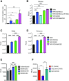
Fig 1. ACE2-independent SARS-CoV-2 entry in macrophages.
(A-B) Representative ACE2 mRNA expression (A) and flow cytometry profiles showing ACE2 surface expression (B) of HEK293T cells stably expressing ACE2 and primary MDMs from multiple donors. (C-D) Parental (vector) and transduced (ACE2) HEK293T cells (C) and primary MDMs from 3 donors (D) were infected with S-pseudotyped lentivirus (20 ng based on p24Gag), in the absence or presence of cathepsin inhibitor (E64D) or TMPRSS2 inhibitor (Camostat), and infection was quantified by measuring luciferase activity at day 3 post-infection. Mock: no virus added, DMSO: no-treatment. The means ± SEM from at least 3 independent experiments are shown. P-values: paired t-test, two-tailed comparing to vector control (C), or one-way ANOVA followed by the Dunnett’s post-test comparing to DMSO (D). ****: p < 0.0001.
Fig 2
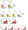
Fig 2. CD169 is a SARS-CoV-2 attachment and entry factor in macrophages.
(A) Binding of SARS-CoV-2 S protein (Wuhan isolate) to THP1 monocytes expressing wt CD169, mutant CD169 (R116A), ACE2, or both wt CD169 and ACE2 (CD169/ACE2). (B) THP1/PMA macrophages were infected with Wuhan (B), Delta (C) or Omicron (D) S-pseudotyped lentivirus (20 ng p24Gag). Infection was quantified by measuring luciferase activity at 3 dpi. Relative light units (RLUs) from each cell line were normalized to no virus control (mock). The means ± SEM are shown from at least five independent experiments. (E) Primary MDMs from three donors overexpressing either CD169 or ACE2, or vector control were infected with Wuhan S-pseudotyped lentivirus (20 ng p24Gag) for 3 days, followed by analysis of luciferase activity in whole cell lysates. (F) Untransduced primary MDMs (representative of 3 donors) were pre-treated with anti-CD169 mAb (20 μg/ml, 7D2), IgG1, or left untreated (NT) for 30 min at 4°C prior to infection with Wuhan S-pseudotyped lentivirus (20 ng p24Gag) for 3 days, followed by analysis of luciferase activity. RLUs from each donor in each group were normalized to no virus control (mock). The means ± SEM from 3 independent experiments are shown. P-values: paired t-test, two-tailed (A), one-way ANOVA followed by the Dunnett’s post-test (B) or Tukey’s post-test comparing to untransduced cells (C-E), or each pre-treatment condition (F). **: p < 0.01; ***: p < 0.001; ****: p < 0.0001; ns: not significant.
Fig 3
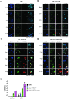
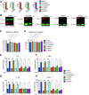
Fig 3. SARS-CoV-2 establishes abortive infection in CD169+ THP1/PMA macrophages.
(A-D) Representative immunofluorescence images (100x) of THP1/PMA macrophages infected with SARS-CoV-2 (MOI = 1) and stained for nucleus (DAPI, blue), SARS-CoV-2 dsRNA (green), and SARS-CoV-2 nucleocapsid (N, red), at indicated times post infection. Images shown for each cell line; untransduced control (parental, A), CD169+ (B), ACE2+ (C), and CD169+/ACE2+ (D). Bar = 25 μm. (E) Culture supernatants from SARS-CoV-2 infected THP1/PMA macrophages were harvested at 24 hpi and viral titers determined by TCID50 assay. The means ± SEM from 3 independent experiments are shown.
Fig 4
Fig 4. CD169-mediated SARS-CoV-2 infection results in restricted viral RNA expression in THP1/PMA macrophages.
(A) Single molecule RNA FISH analysis of viral positive sense gRNA using high fidelity probes in SARS-CoV-2 infected THP1/PMA macrophages (MOI = 10) at the indicated timepoints. Representative fields of cells were hybridized at indicated times with 7 sets of smFISH probes labeled with Quasar670 targeting the + strand of SARS-CoV-2 ORF1a (NSP1-3) and N transcripts. Data are representative of 2 independent experiments. Bar = 50 μm. (B) smFISH analysis of viral RNAs using ORF1a specific probe set (labeled with Texas Red) and N specific probe set (labelled with TMR) in SARS-CoV-2 infected THP1/PMA cells (MOI = 10, 24 hpi). Data are representative of 2 independent experiments. Bar = 25 μm. (C) Percentage of infected cells based on the presence of SARS-CoV-2 RNAs determined from 10–20 independent fields (representative field shown in B). (D-G) THP1/PMA macrophages infected with SARS-CoV-2 (MOI = 1) in the absence (D-E) or presence (F-G) of remdesivir (RDV, 1 μM). Total RNA was harvested at indicated times post-infection and expression of viral transcripts was quantified by RT-qPCR. Replication kinetics of SARS-CoV-2 were quantified by (D) total N RNA amplification (values from each group normalized to mock (uninfected) or (E) negative sense antigenome from strand-specific reverse-transcription and ORF1b amplification (mean Ct values for each condition). Expression of total N RNA (F) and E sgRNA (G) transcripts in THP1/PMA macrophages was analyzed at 24 hpi. Values from each group normalized to mock (uninfected). The means ± SEM are shown from at least 3 independent experiments. Significant differences between groups were determined by one-way ANOVA followed by the Dunnett’s post-test (C, F-G), comparing to parental THP1 cells. P-values: *<0.1; **<0.01; ***<0.001; ****<0.0001.
Fig 5
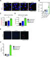
Fig 5. CD169-mediated virus entry leads to restricted SARS-CoV-2 infection in primary MDMs.
(A) Expression of SARS-CoV-2 genomic RNA in primary MDMs imaged using smFISH probes against Orf1a (NSP 1–3), and DAPI for nuclear staining. Data are a representative of 2 independent experiments. Bar = 25 μm. (B) Quantitation of mean fluorescence intensities (MFI) based on the presence of SARS-CoV-2 gRNAs determined from 10–20 independent fields. Significant differences between the groups were determined by one-way ANOVA showing mean fluorescence intensity ± SEM for each group. (C-D) Total RNA from SARS-CoV-2 infected primary untransduced MDMs (MOI = 1, 24 hpi), or MDMs overexpressing either CD169 or ACE2 was analyzed by RT-qPCR for expression of total N RNA (C), and envelope sgRNA (D). Fold expression normalized to mock (uninfected) control for each donor. Significant differences between groups were determined by one-way ANOVA followed by the Dunnett’s post-test (C-D), comparing to untransduced control for each donor. P-values: *<0.1; **<0.01; ****<0.0001. (E) Representative immunofluorescence images (20x) from primary untransduced MDMs, or MDMs overexpressing either CD169 or ACE2, infected with SARS-CoV-2 (MOI = 1, 24 hpi) and stained for nucleus (DAPI, blue), and SARS-CoV-2 N (red). Bar = 25 μm. (F) Culture supernatants from SARS-CoV-2 infected primary MDMs from multiple donors were harvested at 24 hpi and viral titers determined by TCID50 assay.
Fig 6
Fig 6. Restricted de novo expression of SARS-CoV-2 RNA induces pro-inflammatory responses in non-productively infected THP-1/PMA macrophages.
(A-B) PMA-differentiated THP1 cells from parental (THP1) or those expressing CD169, ACE2, or CD169/ACE2 were infected with SARS-CoV-2 (MOI = 1, 24 hpi) in the absence or presence of RDV (1 μM), and total RNA was analyzed by RT-qPCR for pro-inflammatory cytokine and ISG expression. Values were normalized to mock-infected control for each group. Fold-induction for indicated pro-inflammatory cytokines (A, top panel) and ISGs (A, bottom panel) in the absence of RDV, or in the presence of RDV (B). (C-F) Kinetics of pro-inflammatory cytokine/ISG mRNA expression in the absence of RDV in parental (C), CD169-expressing (D), ACE2-expressing (E), and CD169/ACE2-expressing (F) THP1/PMA cells. The means ± SEM are shown from at least 3 independent experiments. Significant differences between groups were determined by one-way ANOVA followed by the Dunnett’s post-test (A-B), comparing to control parental THP1 cells. P-values: *<0.1; **<0.01; ***<0.001; ****<0.0001; ns: not significant.
Fig 7
Fig 7. Endosomal entry of SARS-CoV-2 in MDMs facilitates restricted SARS-CoV-2 RNA synthesis and induction of pro-inflammatory cytokines.
(A-C) MDMs from multiple donors were infected with SARS-CoV-2 (MOI = 1) in the absence or presence of camostat, E64D, or RDV (1 μM). Cells were harvested at 24 hpi and total RNA analyzed by RT-qPCR for expression of viral N RNA (A) and envelope sgRNA (B). (C) Induction of proinflammatory cytokines normalized to mock (uninfected) control from each donor. (D-F) MDMs were infected with SARS-CoV-2 (MOI = 1), in the absence or presence of RDV (1 μM), and viral replication kinetics analyzed by RT-qPCR at indicated timepoints for total N RNA (D) and E sgRNA (E). Induction of indicated cytokine mRNAs (F) normalized to mock from each donor. The means ± SEM are shown from 3 independent experiments, and each symbol represents an independent donor. Significant differences between conditions were determined by one-way ANOVA followed by the Dunnett’s post-test (A-C), or Tukey’s multiple comparisons test (F), comparing to DMSO-treated control. P-values: *<0.1; **<0.01; ***<0.001; ****<0.0001; ns: not significant.
Fig 8
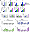
Fig 8. Viral RNA sensing by RIG-I and MDA5 is required for SARS-CoV-2 induced innate immune responses in macrophages.
(A-B) CD169-expressing THP1 monocytes were transduced with lentiviral vectors expressing shRNA against non-specific control (scramble), or specific shRNA sequences against STING, UNC93B1, MAVS, RIG-I, and MDA5. Knockdown of host proteins targeted by shRNAs compared to scramble analyzed by RT-qPCR (A), and immune blotting (B). (C-H) Total RNA from CD169+ THP1/PMA macrophages infected with SARS-CoV-2 (MOI = 1, 24 hpi) was analyzed by RT-qPCR for nucleocapsid (gRNA) (C), envelope (sgRNA, D), and pro-inflammatory cytokines/ISGs, IL6, TNFα, IL-1β and IFNλ1 (E-F), normalized to mock-infected controls for each group. Each knockdown is represented by different colors as in A. The means ± SEM from 3 independent experiments are shown, and significant differences were determined by one-way ANOVA followed by the Dunnett’s post-test comparing to scramble THP1 (C-D), or by two-way ANOVA followed by Bonferroni post-test comparing Mock to SARS-CoV-2 infected in each group (E-H). P-values: *<0.1; **<0.01; ***<0.001; ****<0.0001); ns: not significant.
Fig 9
Fig 9. MAVS is essential for SARS-CoV-2 RNA-induced inflammatory responses in macrophages.
(A) Parental, CD169+, ACE2+, or CD169+/ACE2+ THP1 monocytes were transduced with lentiviral vectors expressing shRNA against scrambled sequence (-), or MAVS sequence (+), and knockdown of MAVS in each cell line was confirmed by western blotting (A). (B-C) MAVS-deficient THP1/PMA macrophages were infected with SARS-CoV-2 (MOI = 1, 24 hpi), and total RNA analyzed by RT-qPCR for (B) viral transcripts (nucleocapsid), and (C) IL6, TNFα, IL-1β and IFNλ1 mRNA expression, normalized to mock-infected controls for each group. The means ± SEM from 3 independent experiments are shown, and significant differences were determined by one-way ANOVA followed by the Tukey-Kramer post-test within groups (B, and C), or Dunnett’s post-test between groups (C). P-values: *<0.05; **<0.01; ***<0.001; ****<0.0001; ns: not significant.

Update of

Similar articles

Cited by

References

    1. Del Valle DM, Kim-Schulze S, Huang HH, Beckmann ND, Nirenberg S, Wang B, et al.. An inflammatory cytokine signature predicts COVID-19 severity and survival. Nat Med. 2020;26: 10. doi: 10.1038/s41591-020-1051-9 - DOI - PMC - PubMed
    1. Zhou F, Yu T, Du R, Fan G, Liu Y, Liu Z, et al.. Clinical course and risk factors for mortality of adult inpatients with COVID-19 in Wuhan, China: a retrospective cohort study. Lancet. 2020;395: 10229. doi: 10.1016/S0140-6736(20)30566-3 - DOI - PMC - PubMed
    1. Chen G, Wu D, Guo W, Cao Y, Huang D, Wang H, et al.. Clinical and immunological features of severe and moderate coronavirus disease 2019. J Clin Invest. 2020;130: 5. doi: 10.1172/JCI137244 - DOI - PMC - PubMed
    1. Ruan Q, Yang K, Wang W, Jiang L, Song J. Clinical predictors of mortality due to COVID-19 based on an analysis of data of 150 patients from Wuhan, China. Intensive Care Med. 2020;46: 5. doi: 10.1007/s00134-020-05991-x - DOI - PMC - PubMed
    1. Pugin J, Ricou B, Steinberg KP, Suter PM, Martin TR. Proinflammatory activity in bronchoalveolar lavage fluids from patients with ARDS, a prominent role for interleukin-1. Am J Respir Crit Care Med. 1996;153: 6 Pt 1. doi: 10.1164/ajrccm.153.6.8665045 - DOI - PubMed

Publication types