

Construction of Artificial Ovaries with Decellularized Porcine Scaffold and Its Elicited Immune Response after Xenotransplantation in Mice
- PMID: 36278634
- PMCID: PMC9589985
- DOI: 10.3390/jfb13040165
Construction of Artificial Ovaries with Decellularized Porcine Scaffold and Its Elicited Immune Response after Xenotransplantation in Mice
Abstract
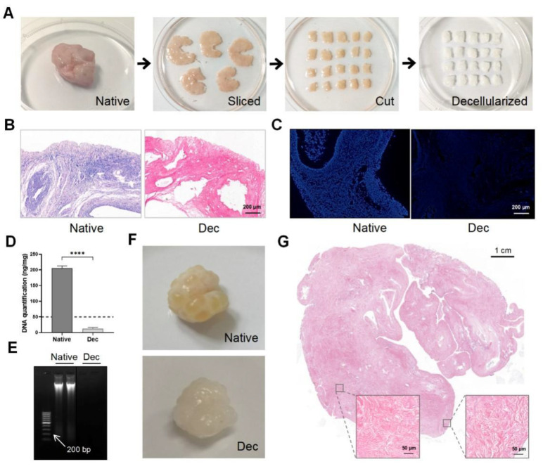
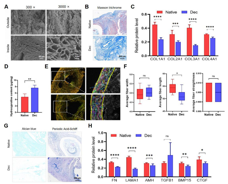
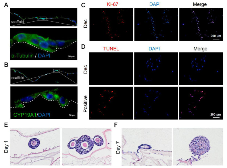
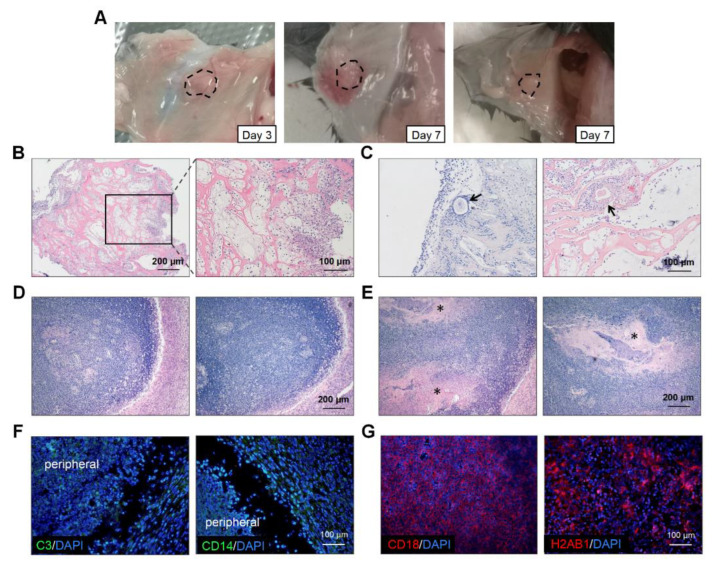
Substitution by artificial ovary is a promising approach to restore ovarian function, and a decellularized extracellular matrix can be used as a supporting scaffold. However, biomimetic ovary fabrication and immunogenicity requires more investigation. In this study, we proposed an effective decellularization protocol to prepare ovarian scaffolds, which were characterized by few nuclear substances and which retained the extracellular matrix proteins. The ovarian tissue shape and 3-dimensional structure were well-preserved after decellularization. Electron micrographs demonstrated that the extracellular matrix fibers in the decellularized group had similar porosity and structure to those of native ovaries. Semi-quantification analysis confirmed that the amount of extracellular matrix proteins was reduced, but the collagen fiber length, width, and straightness did not change significantly. Granulosa cells were attached and penetrated into the decellularized scaffold and exhibited high proliferative activity with no visible apoptotic cells on day 15. Follicle growth was compromised on day 7. The implanted artificial ovaries did not restore endocrine function in ovariectomized mice. The grafts were infiltrated with immune cells within 3 days, which damaged the artificial ovary morphology. The findings suggest that immune rejection plays an important role when using artificial ovaries.
Keywords: artificial ovaries; decellularization; immune response; ovarian function.
Conflict of interest statement
The authors declare no conflict of interest.
Figures

Similar articles
-
Extracellular matrix-derived scaffolds in constructing artificial ovaries for ovarian failure: a systematic methodological review.Hum Reprod Open. 2023 Apr 20;2023(2):hoad014. doi: 10.1093/hropen/hoad014. eCollection 2023. Hum Reprod Open. 2023. PMID: 37180603 Free PMC article. Review.
-
Decellularized human ovarian scaffold based on a sodium lauryl ester sulfate (SLES)-treated protocol, as a natural three-dimensional scaffold for construction of bioengineered ovaries.Stem Cell Res Ther. 2018 Sep 26;9(1):252. doi: 10.1186/s13287-018-0971-5. Stem Cell Res Ther. 2018. PMID: 30257706 Free PMC article.
-
Initial steps in reconstruction of the human ovary: survival of pre-antral stage follicles in a decellularized human ovarian scaffold.Hum Reprod. 2019 Aug 1;34(8):1523-1535. doi: 10.1093/humrep/dez077. Hum Reprod. 2019. PMID: 31286144
-
Liquid nitrogen improves the decellularization effectiveness of whole-ovary.Cryo Letters. 2024 May-Jun;45(3):177-184. Cryo Letters. 2024. PMID: 38709189
-
Comparing various protocols of human and bovine ovarian tissue decellularization to prepare extracellular matrix-alginate scaffold for better follicle development in vitro.BMC Biotechnol. 2021 Jan 20;21(1):8. doi: 10.1186/s12896-020-00658-3. BMC Biotechnol. 2021. PMID: 33472624 Free PMC article.
Cited by
-
Extracellular matrix-derived scaffolds in constructing artificial ovaries for ovarian failure: a systematic methodological review.Hum Reprod Open. 2023 Apr 20;2023(2):hoad014. doi: 10.1093/hropen/hoad014. eCollection 2023. Hum Reprod Open. 2023. PMID: 37180603 Free PMC article. Review.
-
Perfusion and Ultrasonication Produce a Decellularized Porcine Whole-Ovary Scaffold with a Preserved Microarchitecture.Cells. 2023 Jul 15;12(14):1864. doi: 10.3390/cells12141864. Cells. 2023. PMID: 37508528 Free PMC article.
References
Grants and funding
LinkOut - more resources
Full Text Sources
