. 2022 Oct 7:13:1002898.
doi: 10.3389/fimmu.2022.1002898. eCollection 2022.

B7-H3-targeting Fc-optimized antibody for induction of NK cell reactivity against sarcoma

Affiliations

B7-H3-targeting Fc-optimized antibody for induction of NK cell reactivity against sarcoma

Ilona Hagelstein et al. Front Immunol. .

Abstract

Natural killer (NK) cells largely contribute to antibody-dependent cellular cytotoxicity (ADCC), a central factor for success of monoclonal antibodies (mAbs) treatment of cancer. The B7 family member B7-H3 (CD276) recently receives intense interest as a novel promising target antigen for immunotherapy. B7-H3 is highly expressed in many tumor entities, whereas expression on healthy tissues is rather limited. We here studied expression of B7-H3 in sarcoma, and found substantial levels to be expressed in various bone and soft-tissue sarcoma subtypes. To date, only few immunotherapeutic options for treatment of sarcomas that are limited to a minority of patients are available. We here used a B7-H3 mAb to generate chimeric mAbs containing either a wildtype Fc-part (8H8_WT) or a variant Fc part with amino-acid substitutions (S239D/I332E) to increase affinity for CD16 expressing NK cells (8H8_SDIE). In comparative studies we found that 8H8_SDIE triggers profound NK cell functions such as activation, degranulation, secretion of IFNγ and release of NK effector molecules, resulting in potent lysis of different sarcoma cells and primary sarcoma cells derived from patients. Our findings emphasize the potential of 8H8_SDIE as novel compound for treatment of sarcomas, particularly since B7-H3 is expressed in bone and soft-tissue sarcoma independent of their subtype.

Keywords: B7-H3; Fc-optimized; NK cells; immunotherapy; mAb; sarcoma.


Conflict of interest statement

GJ, HS, LZ, and TM are listed as inventors on the patent application “Antibodies targeting, and other modulators of, the CD276 antigen, and uses thereof,” EP3822288A1, applicant German Cancer Research Center, Heidelberg, Germany. The remaining authors declare that the research was conducted in the absence of any commercial or financial relationships that could be construed as a potential conflict of interest.

Figures
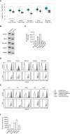
Figure 1
Figure 1
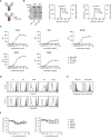
Characterization of B7-H3 expression in sarcoma cell lines and patient-derived sarcoma cells. (A) Relative mRNA expression of B7-H3 in indicated tumor and corresponding normal tissues was analyzed using the online web server GEPIA. T, tumor tissue; N, normal tissue (B) B7-H3 mRNA expression of sarcoma cell lines RD-ES, SaOs, SW1353, SW872 and SW982 was determined via RT-PCR with GAPDH serving as control. PCR products were visualized by agarose gel electrophoresis. (C) mRNA expression analysis of B7-H3 mRNA relative to GAPDH mRNA in five different sarcoma cell lines. Results for n=3 experiments are shown. (D, E) Surface B7-H3 expression on the indicated sarcoma cells was analyzed by flow cytometry using mAb against B7-H3 (shaded peaks) and corresponding isotype control (open peaks). Exemplary histograms (upper panels) and dot plots (lower panels) from one representative experiment of a total of three with similar results are shown. B7-H3 expression of (D) sarcoma cell lines and (E) patient-derived sarcoma cells of different subtypes dissociated from primary tumor samples are shown, respectively. (F) B7-H3 molecule counts on sarcoma cell lines were determined by FACS. Results for two independent experiments are shown.
Figure 2
Figure 2
Generation and binding characteristics of B7-H3 specific antibodies. (A) Schematic illustration of the generated B7-H3 specific antibodies, either with wildtype Fc part (8H8_WT) (top) or Fc optimized part to enhance affinity to CD16 (8H8_SDIE) (bottom). Created with BioRender.com. (B) Exemplary results of an SDS PAGE (left panel) for both B7-H3 antibodies and of size exclusion chromatography for 8H8_WT (middle panel) and 8H8_SDIE (right panel) (C) Sarcoma cell lines were incubated with the indicated concentrations of 8H8_WT, 8H8_SDIE or the corresponding isotype controls followed by an anti-human PE conjugate and analyzed by flow cytometry. Exemplary data for mean fluorescence intensity (MFI) levels from one representative experiment of a total of three with similar results are shown. (D) Binding of 8H8_WT and 8H8_SDIE or the corresponding controls (1 µg/ml) to the surface of patient-derived sarcoma cells was analyzed by flow cytometry using the 8H8 antibodies (shaded peaks) and the corresponding isotype controls (open peaks), respectively. (E) Specific binding of 8H8 mAbs to CD16 on NK cells was analyzed by flow cytometry using NK cells (CD3- CD56+) within healthy donor PBMC incubated without (open peaks) or with the 8H8 mAbs (shaded peaks) followed by an anti-human PE conjugate. (F) Sarcoma cells RD-ES, SaOs, SW1353, SW872 and SW982 were incubated with indicated concentrations of 8H8_WT, 8H8_SDIE or the corresponding controls for 24 h (left panel) or 72 h (right panel), respectively. Then, cells were washed and reincubated with 1 μg/ml of 8H8_SDIE, followed by an anti-human PE conjugate (1:100) and then analyzed by flow cytometry. Relative surface expression of B7-H3 was calculated by defining the mean fluorescence intensity of cells preincubated without antibody as 100%. Exemplary data from one representative experiment of a total of three are shown.
Figure 3
Figure 3
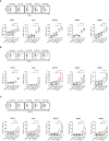
Induction of NK cell reactivity by B7-H3 antibodies against sarcoma cells. PBMC of healthy donors were cultured with or without sarcoma cells at an E:T ratio of 2.5:1 in the presence or absence of B7-H3 antibodies or the corresponding isotype controls (1 μg/mL). (A) Activation of NK cells was determined by expression of CD69 after 24h. In the upper panels, exemplary flow cytometry results obtained with SaOs and in the lower panels data for sarcoma cell lines RD-ES, SaOs, SW1353, SW872 and SW982 (n=5) with PBMC of 4 different donors are shown. (B) Activation of NK cells was determined by expression of CD25 after 72h. In the upper panels exemplary flow cytometry results obtained with RD-ES and in the lower panels data with sarcoma cell lines RD-ES, SaOs, SW1353, SW872 and SW982 with PBMC of 4 different donors are shown. (C) Degranulation of NK cells was determined by expression of CD107a after 4h. In the upper panels exemplary flow cytometry results obtained with SW872 and in the lower panels data with sarcoma cell lines RD-ES, SaOs, SW1353, SW872 and SW982 with PBMC of four independent donors are shown. ns, not significant; *statistically significant differences (p-value < 0.05).
Figure 4
Figure 4
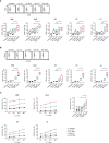
Induction of immunoregulatory molecules by B7-H3 antibodies against sarcoma cells. Healthy donor PBMC were cultured with sarcoma cells at an E:T ratio of 2.5:1 in the presence or absence of 8H8_WT, 8H8_SDIE or the corresponding isotype controls (1 μg/mL) (A,B) Supernatants and NK cells within PBMC were analyzed for IFNγ. (A) Release of IFNγ after 24 h was analyzed in supernatants of cocultures by Legendplex assays. In the left panel, exemplary results with SW1353 are shown, in the right panel results obtained with sarcoma cell lines RD-ES, SaOs, SW1353 and SW872 and with PBMC of four independent donors are shown. (B) Intracellular expression of IFNγ in NK cells within PBMC identified by counterstaining with CD3- CD56+ was analyzed after 4 h by flow cytometry. In the upper panel, exemplary results obtained with SaOs and in the lower panel data obtained with sarcoma cells (RD-ES, SaOs, SW1353, SW872 and SW982) and with PBMC of four independent donors are shown. (C) Supernatants were analyzed for effector molecules Granzyme A, Granzyme B, Perforin, Granulysin and sFasL and for release of immunoregulatory molecules TNFα, IL-2, IFNγ, IL-4, IL-10 after 4 h by Legendplex assays. Shown are pooled results with sarcoma cell lines SW1353 and SW872 and with PBMC of two independent donors. ns, not significant; *statistically significant differences (p-value < 0.05).
Figure 5
Figure 5
Induction of target cell lysis by Fc optimized B7-H3 antibody. PBMC of healthy donors were incubated with different sarcoma cell lines (n = 5) and treated without or with indicated B7-H3 antibodies or corresponding isotype controls (1 μg/mL). (A) Lysis of sarcoma cell lines RD-ES, SaOs, SW1353, SW872 and SW982 (n=5) was analyzed by 2 h Europium cytotoxicity assays. In the top panel, exemplary data obtained with SW982 and one PBMC donor with different E:T ratios and in the bottom panel, pooled data for each cell line obtained with PBMC of healthy donors (n=5) at an E:T ratio of 20:1 are shown. (B) Lysis of sarcoma cell lines RD-ES, SaOs, SW1353, SW872 and SW982 (n=5) was determined after 72 h by flow cytometry-based lysis assays at an E:T ratio of 10:1. In the top panel exemplary dot plots with SW872 and one PBMC donor are shown, the bottom panel depicts combined results for each cell line with PBMC (n=5) of healthy donors. (C) Cell death of sarcoma cells was determined using a live cell imaging system. Sarcoma cell lines RD-ES, SaOs, SW1353, SW872 and SW982 were incubated with PBMC of two healthy donors at an E:T ratio of 10:1 for 120h. T=0 h corresponds to a confluence of 100%. Results are shown as mean ± SD. ns, not significant; *statistically significant differences (p-value < 0.05).
Figure 6
Figure 6
8H8_SDIE induces NK cell reactivity and cytotoxicity against patient-derived sarcoma cells. PBMC of healthy donors were incubated with patient-derived sarcoma cells of different subtypes and treated with the indicated B7-H3 antibodies or the corresponding isotype controls (1 μg/mL). (A) Activation of NK cells was determined by expression of CD69 after 24h. In the top panels, exemplary flow cytometry results obtained with patient-derived synovialsarcoma cells and one PBMC donor and in the bottom panel, combined data with patient-derived sarcoma cells (n=5) and with PBMC of four different donors (n=4) are shown. (B) Degranulation of NK cells was determined by expression of CD107a after 4h. In the upper panels, exemplary flow cytometry results obtained with patient-derived rhabdomyosarcoma cells and one PBMC donor and in the lower panel, data with patient-derived sarcoma cells (n=5) and with PBMC of four independent donors (n=4) are shown. (C) Killing of patient-derived sarcoma cells (n=4) was analyzed by 2 h Europium cytotoxicity assays. In the left panels, exemplary data obtained with PBMC of one healthy donor and patient-derived rhabdomyosarcoma, osteosarcoma, synovialsarcoma and chondrosarcoma cells as indicated with different E:T ratios and on the right, pooled data obtained with PBMC of healthy donors (n=2) and patient-derived sarcoma cells (n=4) at an E:T ratio of 20:1 are shown. ns, not significant; *statistically significant differences (p-value < 0.05).

Similar articles

Cited by

References

    1. Choi JH, Ro JY. The 2020 WHO classification of tumors of bone: An updated review. Adv Anat Pathol (2021) 28(3):119–38. doi: 10.1097/PAP.0000000000000293 - DOI - PubMed
    1. Choi JH, Ro JY. The 2020 WHO classification of tumors of soft tissue: Selected changes and new entities. Adv Anat Pathol (2021) 28(1):44–58. doi: 10.1097/PAP.0000000000000284 - DOI - PubMed
    1. Brown HK, Schiavone K, Gouin F, Heymann MF, Heymann D. Biology of bone sarcomas and new therapeutic developments. Calcif Tissue Int (2018) 102(2):174–95. doi: 10.1007/s00223-017-0372-2 - DOI - PMC - PubMed
    1. Clark MA, Fisher C, Judson I, Thomas JM. Soft-tissue sarcomas in adults. N Engl J Med (2005) 353(7):701–11. doi: 10.1056/NEJMra041866 - DOI - PubMed
    1. Grunewald TG, Alonso M, Avnet S, Banito A, Burdach S, Cidre-Aranaz F, et al. . Sarcoma treatment in the era of molecular medicine. EMBO Mol Med (2020) 12(11):e11131. doi: 10.15252/emmm.201911131 - DOI - PMC - PubMed

Publication types