

Regulatory role of KCa3.1 in immune cell function and its emerging association with rheumatoid arthritis
- PMID: 36275686
- PMCID: PMC9580404
- DOI: 10.3389/fimmu.2022.997621
Regulatory role of KCa3.1 in immune cell function and its emerging association with rheumatoid arthritis
Abstract
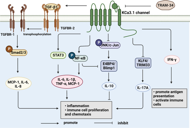
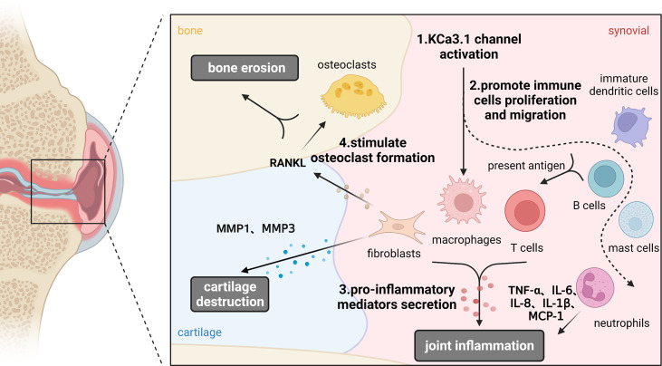
Rheumatoid arthritis (RA) is a common autoimmune disease characterized by chronic inflammation. Immune dysfunction is an essential mechanism in the pathogenesis of RA and directly linked to synovial inflammation and cartilage/bone destruction. Intermediate conductance Ca2+-activated K+ channel (KCa3.1) is considered a significant regulator of proliferation, differentiation, and migration of immune cells by mediating Ca2+ signal transduction. Earlier studies have demonstrated abnormal activation of KCa3.1 in the peripheral blood and articular synovium of RA patients. Moreover, knockout of KCa3.1 reduced the severity of synovial inflammation and cartilage damage to a significant extent in a mouse collagen antibody-induced arthritis (CAIA) model. Accumulating evidence implicates KCa3.1 as a potential therapeutic target for RA. Here, we provide an overview of the KCa3.1 channel and its pharmacological properties, discuss the significance of KCa3.1 in immune cells and feasibility as a drug target for modulating the immune balance, and highlight its emerging role in pathological progression of RA.
Keywords: KCa3.1; immune cells; joint inflammation; rheumatoid arthritis; synovitis.
Copyright © 2022 Lin, Zhao, Zhang, Hao, Zhu, Wang, Hu and Zhou.
Conflict of interest statement
The authors declare that the research was conducted in the absence of any commercial or financial relationships that could be construed as a potential conflict of interest.
Figures


Similar articles
-
Protection against cartilage and bone destruction by systemic interleukin-4 treatment in established murine type II collagen-induced arthritis.Arthritis Res. 1999;1(1):81-91. doi: 10.1186/ar14. Epub 1999 Oct 26. Arthritis Res. 1999. PMID: 11056663 Free PMC article.
-
c-Fms-mediated differentiation and priming of monocyte lineage cells play a central role in autoimmune arthritis.Arthritis Res Ther. 2010;12(1):R32. doi: 10.1186/ar2940. Epub 2010 Feb 24. Arthritis Res Ther. 2010. PMID: 20181277 Free PMC article.
-
Functional role of the KCa3.1 potassium channel in synovial fibroblasts from rheumatoid arthritis patients.J Cell Physiol. 2015 Jul;230(7):1677-88. doi: 10.1002/jcp.24924. J Cell Physiol. 2015. PMID: 25545021
-
Acid-Sensing Ion Channel-1a in Articular Chondrocytes and Synovial Fibroblasts: A Novel Therapeutic Target for Rheumatoid Arthritis.Front Immunol. 2021 Jan 28;11:580936. doi: 10.3389/fimmu.2020.580936. eCollection 2020. Front Immunol. 2021. PMID: 33584647 Free PMC article. Review.
-
The p53 status in rheumatoid arthritis with focus on fibroblast-like synoviocytes.Immunol Res. 2021 Jun;69(3):225-238. doi: 10.1007/s12026-021-09202-7. Epub 2021 May 13. Immunol Res. 2021. PMID: 33983569 Review.
Cited by
-
Efficacy of combined tumor irradiation and KCa3.1-targeting with TRAM-34 in a syngeneic glioma mouse model.Sci Rep. 2023 Nov 23;13(1):20604. doi: 10.1038/s41598-023-47552-4. Sci Rep. 2023. PMID: 37996600 Free PMC article.
-
Challenges in the Therapeutic Targeting of KCa Channels: From Basic Physiology to Clinical Applications.Int J Mol Sci. 2024 Mar 4;25(5):2965. doi: 10.3390/ijms25052965. Int J Mol Sci. 2024. PMID: 38474212 Free PMC article. Review.
-
Intracellular acidity impedes KCa3.1 activation by Riluzole and SKA-31.Front Pharmacol. 2024 Apr 4;15:1380655. doi: 10.3389/fphar.2024.1380655. eCollection 2024. Front Pharmacol. 2024. PMID: 38638868 Free PMC article.
-
Regulation of T Lymphocyte Functions through Calcium Signaling Modulation by Nootkatone.Int J Mol Sci. 2024 May 11;25(10):5240. doi: 10.3390/ijms25105240. Int J Mol Sci. 2024. PMID: 38791278 Free PMC article.
-
Exploring the therapeutic opportunities of potassium channels for the treatment of rheumatoid arthritis.Front Pharmacol. 2024 May 9;15:1286069. doi: 10.3389/fphar.2024.1286069. eCollection 2024. Front Pharmacol. 2024. PMID: 38783950 Free PMC article. Review.
References
Publication types
MeSH terms
Substances
LinkOut - more resources
Full Text Sources
Medical
Miscellaneous