

Are there roles for heterogeneous ribosomes during sleep in the rodent brain?
- PMID: 36275625
- PMCID: PMC9582285
- DOI: 10.3389/fmolb.2022.1008921
Are there roles for heterogeneous ribosomes during sleep in the rodent brain?
Abstract
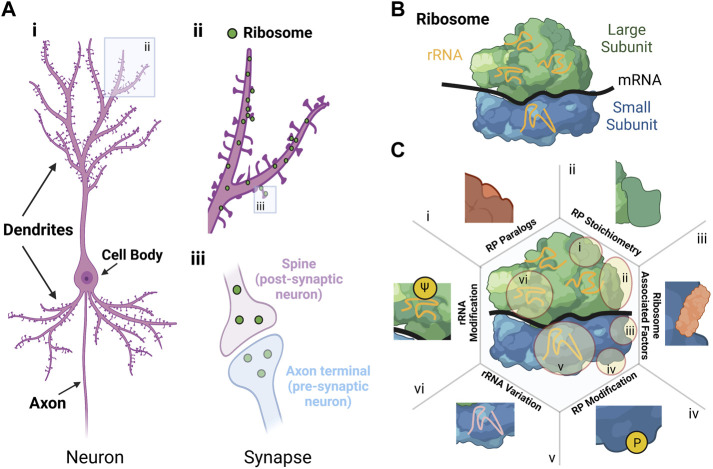
The regulation of mRNA translation plays an essential role in neurons, contributing to important brain functions, such as brain plasticity and memory formation. Translation is conducted by ribosomes, which at their core consist of ribosomal proteins (RPs) and ribosomal RNAs. While translation can be regulated at diverse levels through global or mRNA-specific means, recent evidence suggests that ribosomes with distinct configurations are involved in the translation of different subsets of mRNAs. However, whether and how such proclaimed ribosome heterogeneity could be connected to neuronal functions remains largely unresolved. Here, we postulate that the existence of heterologous ribosomes within neurons, especially at discrete synapses, subserve brain plasticity. This hypothesis is supported by recent studies in rodents showing that heterogeneous RP expression occurs in dendrites, the compartment of neurons where synapses are made. We further propose that sleep, which is fundamental for brain plasticity and memory formation, has a particular role in the formation of heterologous ribosomes, specialised in the translation of mRNAs specific for synaptic plasticity. This aspect of our hypothesis is supported by recent studies showing increased translation and changes in RP expression during sleep after learning. Thus, certain RPs are regulated by sleep, and could support different sleep functions, in particular brain plasticity. Future experiments investigating cell-specific heterogeneity in RPs across the sleep-wake cycle and in response to different behaviour would help address this question.
Keywords: brain plasticity; neurites; neuron; ribosomal protein; ribosome heterogeneity; sleep; synapse.
Copyright © 2022 Buchanan, Smith, Gerber and Seibt.
Conflict of interest statement
The authors declare that the research was conducted in the absence of any commercial or financial relationships that could be construed as a potential conflict of interest.
Figures
Similar articles
-
Hippocampal neurons' cytosolic and membrane-bound ribosomal transcript profiles are differentially regulated by learning and subsequent sleep.Proc Natl Acad Sci U S A. 2021 Nov 30;118(48):e2108534118. doi: 10.1073/pnas.2108534118. Proc Natl Acad Sci U S A. 2021. PMID: 34819370 Free PMC article.
-
On-Site Ribosome Remodeling by Locally Synthesized Ribosomal Proteins in Axons.Cell Rep. 2019 Dec 10;29(11):3605-3619.e10. doi: 10.1016/j.celrep.2019.11.025. Cell Rep. 2019. PMID: 31825839 Free PMC article.
-
Heterogeneous Ribosomes Preferentially Translate Distinct Subpools of mRNAs Genome-wide.Mol Cell. 2017 Jul 6;67(1):71-83.e7. doi: 10.1016/j.molcel.2017.05.021. Epub 2017 Jun 15. Mol Cell. 2017. PMID: 28625553 Free PMC article.
-
Ribosome specialization and its potential role in the control of protein translation and skeletal muscle size.J Appl Physiol (1985). 2019 Aug 1;127(2):599-607. doi: 10.1152/japplphysiol.00946.2018. Epub 2019 Jan 3. J Appl Physiol (1985). 2019. PMID: 30605395 Review.
-
Specialized ribosomes and the control of translation.Biochem Soc Trans. 2018 Aug 20;46(4):855-869. doi: 10.1042/BST20160426. Epub 2018 Jul 9. Biochem Soc Trans. 2018. PMID: 29986937 Review.
Cited by
-
The brain's dark transcriptome: Sequencing RNA in distal compartments of neurons and glia.Curr Opin Neurobiol. 2023 Aug;81:102725. doi: 10.1016/j.conb.2023.102725. Epub 2023 May 15. Curr Opin Neurobiol. 2023. PMID: 37196598 Free PMC article. Review.
References
LinkOut - more resources
Full Text Sources