

Cellular events during ovine implantation and impact for gestation
- PMID: 36249852
- PMCID: PMC9536072
- DOI: 10.21451/1984-3143-AR2018-0014
Cellular events during ovine implantation and impact for gestation
Abstract
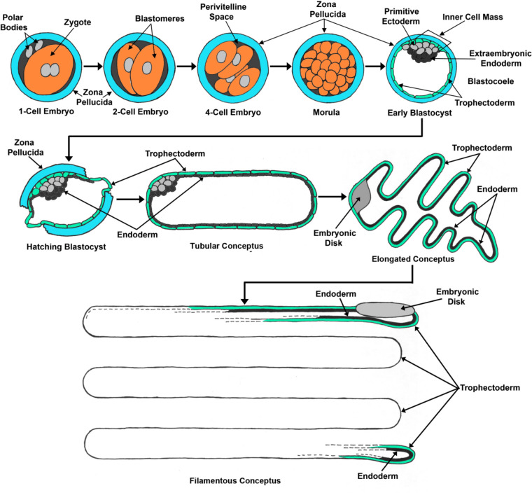
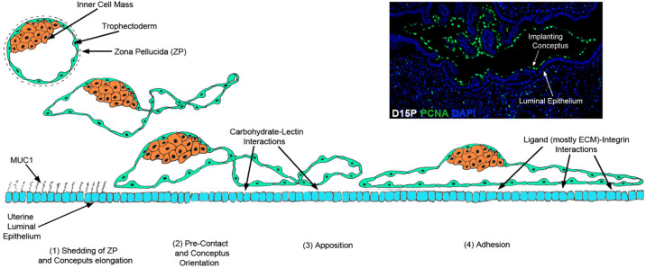
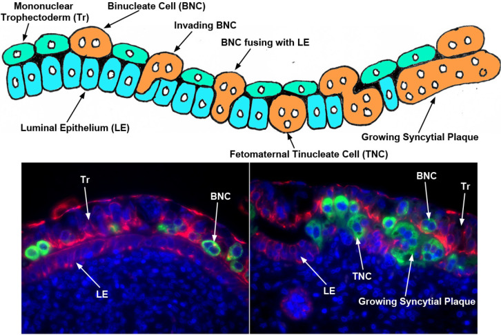
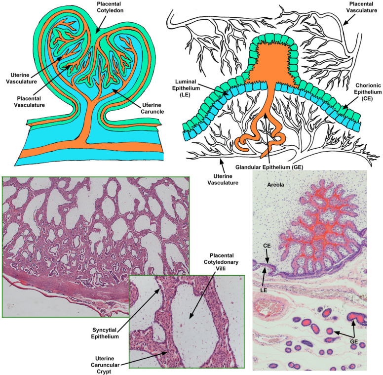
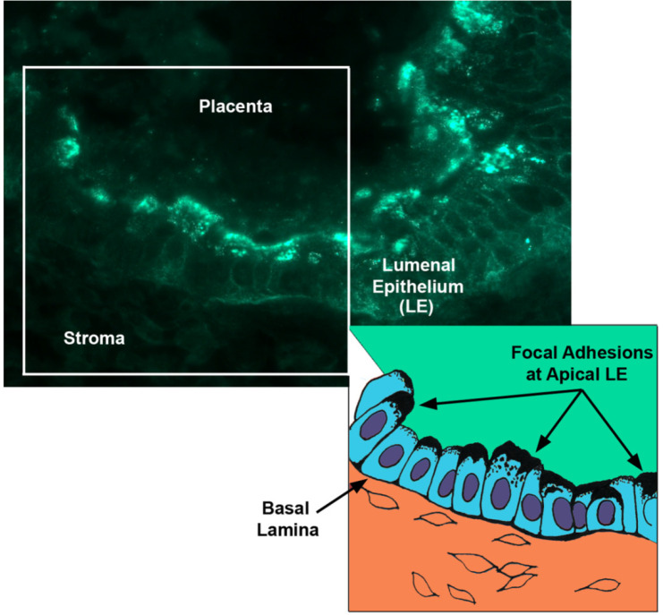
The establishment of pregnancy in sheep includes elongation of the blastocyst into a filamentous conceptus, pregnancy recognition, production of histotroph, attachment of the conceptus to the endometrium for implantation, and development of synepitheliochorial placentation. These processes are complex, and this review describes some of the molecular events that underlie and support successful pregnancy. The free-floating sheep blastocyst elongates into a filamentous conceptus and metabolizes, or is responsive to, molecules supplied by the endometrium as histotroph. Amongst these molecules are SPP1, glucose and fructose, and arginine that stimulate the MTOR nutrient sensing system. The placental trophectoderm of elongating conceptuses initiate pregnancy recognition and implantation. The mononucleate cells of the trophectoderm secrete IFNT, which acts on the endometrial LE to block increases in estrogen receptor α to preclude oxytocin receptor expression, thereby preventing oxytocin from inducing luteolytic pulses of PGF2α. In addition, IFNT increases expression of IFN stimulated genes in the endometrial stroma, including ISG15, a functional ubiquitin homologue. Implantation is the initial step in placentation, and includes sequential pre-contact, apposition, and adhesion phases. Implantation in sheep includes downregulation of Muc1 and interaction of GLYCAM1, galectin 15 (LGALS15) and SPP1 with lectins and integrins (αvβ3). Sheep have synepitheliochorial placentation in which mononucleate trophectoderm cells fuse to form binucleate cells (BNCs). BNCs migrate and fuse with endometrial LE cells to form trinucleate syncytial cells, and these syncytia enlarge through continued BNC fusion to form syncytial plaques that form the interface between endometrial and placental tissues within the placentome. The placentae of sheep organize into placentomal and interplacentomal regions. In placentomes there is extensive interdigitation of endometrial and placental tissues to provide hemotrophic nutrition to the fetus. In interplacentomal regions there is epitheliochorial attachment of endometrial LE to trophectoderm, mediated through focal adhesion assembly, and areolae that take up histotroph secreted by endometrial GE.
Keywords: conceptus; endometrium; placentation; pregnancy; sheep..
Figures

Similar articles
-
Fetal-maternal interactions during the establishment of pregnancy in ruminants.Soc Reprod Fertil Suppl. 2007;64:379-96. doi: 10.5661/rdr-vi-379. Soc Reprod Fertil Suppl. 2007. PMID: 17491160
-
Osteopontin: a leading candidate adhesion molecule for implantation in pigs and sheep.J Anim Sci Biotechnol. 2014 Dec 17;5(1):56. doi: 10.1186/2049-1891-5-56. eCollection 2014. J Anim Sci Biotechnol. 2014. PMID: 25671104 Free PMC article. Review.
-
Review: Implantation and placentation in ruminants.Animal. 2023 May;17 Suppl 1:100796. doi: 10.1016/j.animal.2023.100796. Animal. 2023. PMID: 37567669 Review.
-
Cell-Specific Expression of Enzymes for Serine Biosynthesis and Glutaminolysis in Farm Animals.Adv Exp Med Biol. 2021;1285:17-28. doi: 10.1007/978-3-030-54462-1_2. Adv Exp Med Biol. 2021. PMID: 33770400
-
Biology of progesterone action during pregnancy recognition and maintenance of pregnancy.Front Biosci. 2002 Sep 1;7:d1879-98. doi: 10.2741/spencer. Front Biosci. 2002. PMID: 12161340 Review.
Cited by
-
Early Embryonic Development in Agriculturally Important Species.Animals (Basel). 2024 Jun 26;14(13):1882. doi: 10.3390/ani14131882. Animals (Basel). 2024. PMID: 38997994 Free PMC article. Review.
-
Consequences of twinning induction to Noemi ewes by a recombinant human follicle-stimulating hormone compared with pituitary-derived porcine follicle-stimulating hormone on follicular dynamics, maternal biochemical attributes, and neonatal traits.Vet World. 2019 Apr;13(4):633-641. doi: 10.14202/vetworld.2020.633-641. Epub 2020 Apr 8. Vet World. 2019. PMID: 32546905 Free PMC article.
-
Peroxisome Proliferator-Activated Receptor γ Regulates Lipid Metabolism in Sheep Trophoblast Cells through mTOR Pathway-Mediated Autophagy.PPAR Res. 2023 Nov 8;2023:6422804. doi: 10.1155/2023/6422804. eCollection 2023. PPAR Res. 2023. PMID: 38020065 Free PMC article.
-
Single-nuclei RNA sequencing (snRNA-seq) uncovers trophoblast cell types and lineages in the mature bovine placenta.Proc Natl Acad Sci U S A. 2023 Mar 21;120(12):e2221526120. doi: 10.1073/pnas.2221526120. Epub 2023 Mar 13. Proc Natl Acad Sci U S A. 2023. PMID: 36913592 Free PMC article.
-
Phosphate, calcium, and vitamin D signaling, transport, and metabolism in the endometria of cyclic ewes.J Anim Sci Biotechnol. 2023 Jan 12;14(1):13. doi: 10.1186/s40104-022-00803-2. J Anim Sci Biotechnol. 2023. PMID: 36631878 Free PMC article.
References
-
- Al-Shami R, Sorense, ES, Ek-Rylander B, Andersson G, Carson DD, Farach-Carson MC. Phosphorylated osteopontin promotes migration of human choriocarcinoma cells via a p70S6 kinase- dependent pathway. J Cell Biochem. 2005;94:1218–1233. - PubMed
-
- Aplin JD, Meseguer M, Simon C, Ortiz ME, Croxatto H, Jones CJ. MUC1, glycans and the cell-surface barrier to embryo implantation. Biochem Soc Trans. 2001;29:153–156. - PubMed
-
- Austin KJ, Bany BM, Belden EL, Rempel LA, Cross JC, Hansen TR. Interferon-stimulated gene-15 (Isg15) expression is up-regulated in the mouse uterus in response to the implanting conceptus. Endocrinology. 2004;144:3107–3113. - PubMed
-
- Bazer FW, Johnson GA, Spencer TE. Growth and development: pre-implantation embryo. In: Pond WG, Bell AW, editors. Encyclopedia of Animal Science. Vol. 1. New York, NY: Marcel Dekker; 2005. pp. 1–3.
-
- Bazer FW, Spencer TE, Thatcher WW. Growth and development of the ovine conceptus. J Anim Sci. 2011;90:159–170. - PubMed
LinkOut - more resources
Full Text Sources
Research Materials
Miscellaneous