

Review
doi: 10.3348/jksr.2020.81.1.58.
Epub 2020 Jan 31.
[Diffusion-Weighted Magnetic Resonance Imaging of Spine]
[Article in
Korean]
- PMID: 36238128
- PMCID: PMC9432087
- DOI: 10.3348/jksr.2020.81.1.58
Item in Clipboard
Review
[Diffusion-Weighted Magnetic Resonance Imaging of Spine]
[Article in
Korean]
Taehan Yongsang Uihakhoe Chi.
2020 Jan.
Abstract
In this study, we evaluated the technical characteristics and usefulness of diffusion-weighted magnetic resonance imaging for discrimination between benign and malignant vertebral fractures, for detection and differentiation of multiple myeloma or metastases, and for response monitoring in malignant vertebral lesions after anticancer drug therapy or radiation therapy.
척추영상에 사용되는 확산강조영상의 기술적 측면, 양성과 악성 척추체 압박골절의 구분, 다발성골수종이나 전이암의 발견과 감별진단, 그리고 항암화학요법이나 방사선치료 후 반응 평가에 대해 기술하고자 한다.
Copyrights © 2020 The Korean Society of Radiology.
Conflict of interest statement
Conflicts of Interest: The authors have no potential conflicts of interest to disclose.
Figures
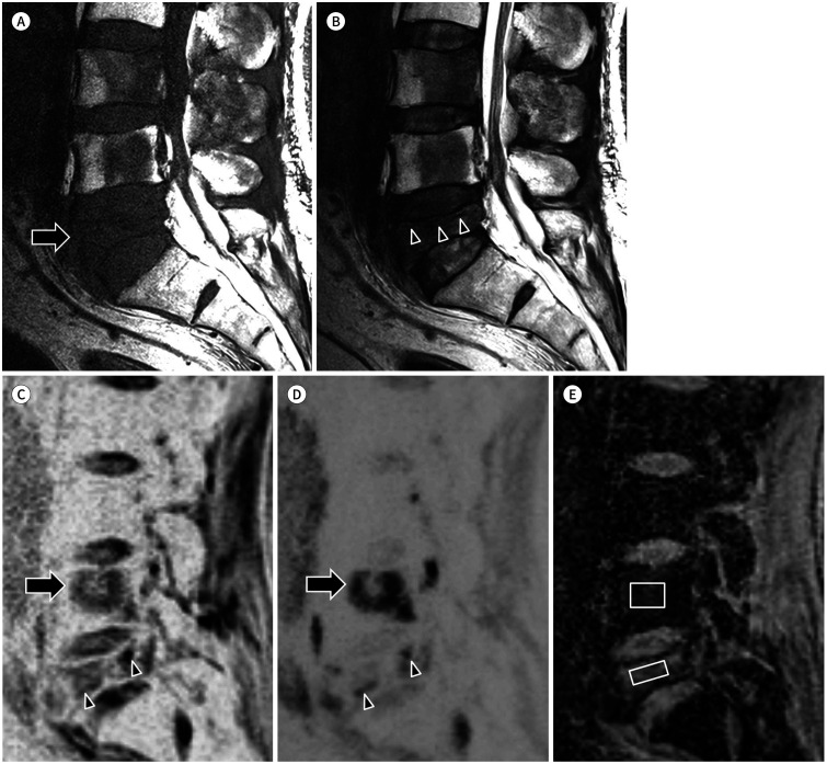
A. T1-weighted sagittal image showing decreased height of the L5 vertebral body (arrow) and low signal intensity lesions in the L3, L4, and L5 vertebral bodies. B. T2-weighted sagittal image showing a band-like low signal (arrowheads) parallel with the upper endplate of the L5 vertebral body. C, D. Diffusion-weighted images of b = 0 (C) and b = 800 (D) showing consistently increased signal in the L4 vertebral body (arrows) and seemingly decreased extent of the high signal area in the L5 vertebral body (arrowheads). E. ADC map showing a low ADC value (0.8 × 10⁻³ mm2/s) of the L4 lesion, indicating a viable metastasis, and a relatively high ADC value (1.5 × 10⁻³ mm2/s) of the L5 lesion, indicating a benign compression fracture. ADC = apparent diffusion coefficient
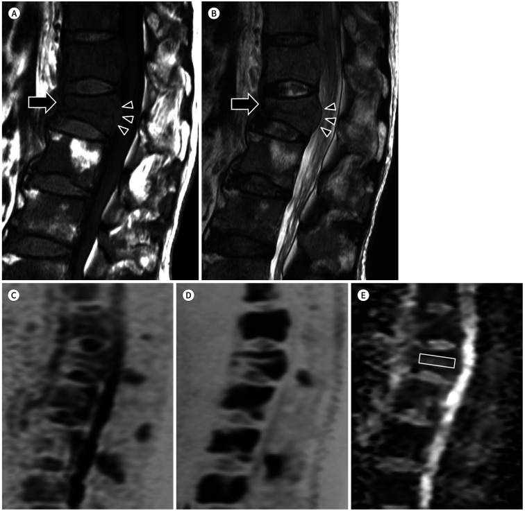
A, B. T1-weighted (A) and T2-weighted (B) images showing areas with heterogeneous low signal in the vertebral bodies and anterior wedging deformity (arrows) with posterior convexity of the T11 vertebral body (arrowheads). C, D. Diffusion-weighted images with b = 0 (C) and b = 800 (D) showing diffusion restriction in multiple thoracolumbar vertebral bodies and spinous processes. E. ADC map showing a low ADC value (0.9 × 10⁻³ mm2/s) of the T11 lesion, indicating a pathologic compression fracture. ADC = apparent diffusion coefficient
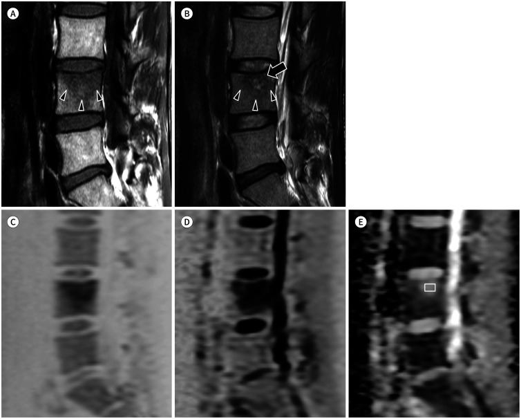
A, B. T1-weighted image (A) showing poorly defined slightly low signal area in the bone marrow of the L4 vertebral body (arrowheads); T2-weighted image (B) showing poorly defined fluid-like high signal area (arrow) with adjacent intermediate-to-low signal area in the bone marrow of the L4 vertebral body (arrowheads). C, D. Diffusion-weighted images of b = 0 (C), showing increased signal area in the upper portion of the L4 vertebral body, and b = 800 (D), showing increased signal intensity (T2 shine-through effect) in the upper portion of the L4 vertebral body. E. ADC map showing a high ADC value (2.3 × 10⁻³ mm2/s) of the L4 lesion, indicating a benign process. ADC = apparent diffusion coefficient
A. T1-weighted image showing an ovoid area with low signal intensity in the L1 vertebral body. B. T2-weighted image showing iso- to slightly low signal intensity in the same area of the L1 vertebral body. C, D. Diffusion-weighted images with b = 0 (C) and b = 800 (D) showing high signal intensity in the area of the L1 vertebral body, indicating diffusion restriction. E. ADC map showing a low ADC value (0.8 × × 10−3mm2/s) of the L1 lesion, indicating a viable bone metastasis. ADC = apparent diffusion coefficient
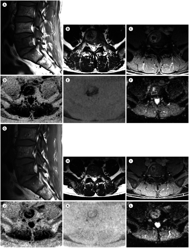
A–C. T1-weighted sagittal (A), T2-weighted axial (B), and post-contrast T1-weighted axial (C) images showing a metastatic bone lesion with enhancement and central necrosis in the L5 vertebral body. D, E. Diffusion-weighted images of b = 0 (D) and b = 1000 (E) showing diffusion restriction in the periphery of the bone lesion. F. ADC map showing a relatively low ADC value (0.7 × × 10−3mm2/s) in the non-necrotic enhancing portion, indicating a viable tumor with high cellularity. G–I. T1-weighted sagittal (G), T2-weighted axial (H) and post-contrast T1-weighted axial (I) images showing decreased extent of the metastatic bone lesion with enhancement and central necrosis in the L5 vertebral body. J–L. Diffusion-weighted images of b = 0 (J) and b = 1000 (K) showing decreased extent of diffusion restriction in the periphery of the bone lesion; ADC map (L) showing an increased ADC value (1.2 × 10⁻³mm2/s) in the non-necrotic enhancing portion, indicating decreased viable tumor burden. ADC = apparent diffusion coefficient
Similar articles
-
Diffusion-weighted MR imaging of bone marrow: differentiation of benign versus pathologic compression fractures.Radiology. 1998 May;207(2):349-56. doi: 10.1148/radiology.207.2.9577479. Radiology. 1998. PMID: 9577479
-
Diffusion-weighted Magnetic Resonance Imaging in Non-traumatic Vertebral Collapse: A Relook Into Its Utility in Making the Diagnosis in a Population Where Infections of Spine Are a Common Cause.J Med Imaging Radiat Sci. 2018 Mar;49(1):90-96. doi: 10.1016/j.jmir.2017.07.001. Epub 2017 Oct 16. J Med Imaging Radiat Sci. 2018. PMID: 30479295
-
Single shot fast spin echo diffusion-weighted MR imaging of the spine; Is it useful in differentiating malignant metastatic tumor infiltration from benign fracture edema?Clin Imaging. 2004 Mar-Apr;28(2):102-8. doi: 10.1016/S0899-7071(03)00247-X. Clin Imaging. 2004. PMID: 15050221
-
Research synthesis: what is the diagnostic performance of magnetic resonance imaging to discriminate benign from malignant vertebral compression fractures? Systematic review and meta-analysis.Spine (Phila Pa 1976). 2012 May 20;37(12):E736-44. doi: 10.1097/BRS.0b013e3182458cac. Spine (Phila Pa 1976). 2012. PMID: 22210011 Review.
-
Diffusion imaging of the vertebral bone marrow.NMR Biomed. 2017 Mar;30(3). doi: 10.1002/nbm.3333. Epub 2015 Jun 26. NMR Biomed. 2017. PMID: 26114411 Review.
Cited by
-
[Imaging Findings of Spinal Metastases with Differential Diagnosis: Focusing on Solitary Spinal Lesion in Older Patients].J Korean Soc Radiol. 2024 Jan;85(1):77-94. doi: 10.3348/jksr.2023.0156. Epub 2024 Jan 26. J Korean Soc Radiol. 2024. PMID: 38362381 Free PMC article. Review. Korean.
References
-
- Chokshi FH, Law M, Gibbs WN. Conventional and advanced imaging of spine oncologic disease, nonoperative post-treatment effects, and unique spinal conditions. Neurosurgery. 2018;82:1–23. - PubMed
-
- Gibbs WN, Nael K, Doshi AH, Tanenbaum LN. Spine oncology: imaging and intervention. Radiol Clin North Am. 2019;57:377–395. - PubMed
-
- Tanenbaum LN. Clinical applications of diffusion imaging in the spine. Magn Reson Imaging Clin N Am. 2013;21:299–320. - PubMed
-
- Dietrich O, Geith T, Reiser MF, Baur-Melnyk A. Diffusion imaging of the vertebral bone marrow. NMR Biomed. 2017;30:e3333 - PubMed
Publication types
LinkOut - more resources
Full Text Sources
