

Role of Mesenchymal Stem Cells and Extracellular Vesicles in Idiopathic Pulmonary Fibrosis
- PMID: 36232511
- PMCID: PMC9569825
- DOI: 10.3390/ijms231911212
Role of Mesenchymal Stem Cells and Extracellular Vesicles in Idiopathic Pulmonary Fibrosis
Abstract
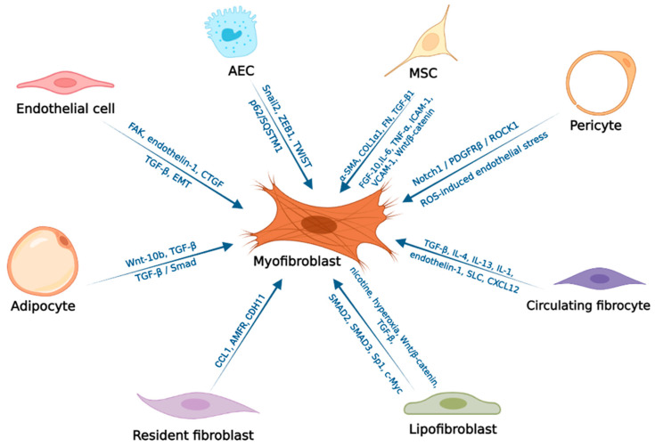
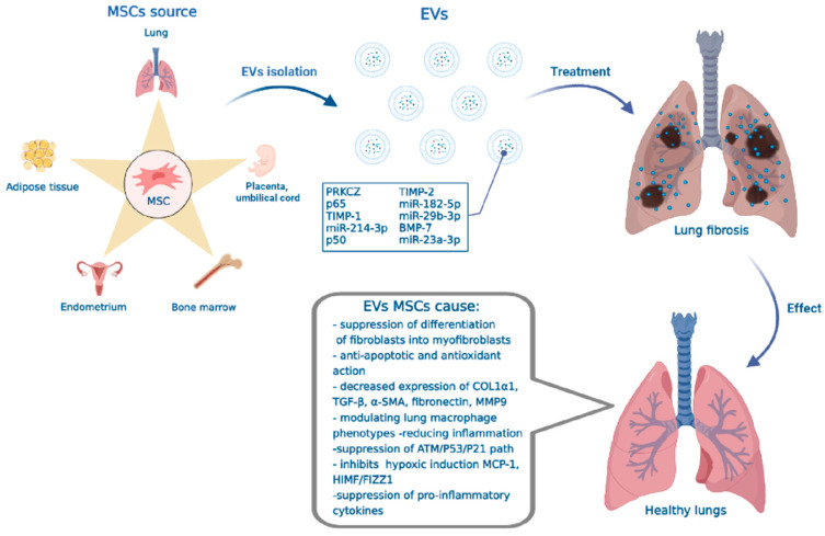
Idiopathic pulmonary fibrosis (IPF) is a progressive interstitial fibrotic disease that leads to disability and death within 5 years of diagnosis. Pulmonary fibrosis is a disease with a multifactorial etiology. The concept of aberrant regeneration of the pulmonary epithelium reveals the pathogenesis of IPF, according to which repeated damage and death of alveolar epithelial cells is the main mechanism leading to the development of progressive IPF. Cell death provokes the migration, proliferation and activation of fibroblasts, which overproduce extracellular matrix, resulting in fibrotic deformity of the lung tissue. Mesenchymal stem cells (MSCs) and extracellular vesicles (EVs) are promising therapies for pulmonary fibrosis. MSCs, and EVs derived from MSCs, modulate the activity of immune cells, inhibit the expression of profibrotic genes, reduce collagen deposition and promote the repair of damaged lung tissue. This review considers the molecular mechanisms of the development of IPF and the multifaceted role of MSCs in the therapy of IPF. Currently, EVs-MSCs are regarded as a promising cell-free therapy tool, so in this review we discuss the results available to date of the use of EVs-MSCs for lung tissue repair.
Keywords: extracellular vesicles; lung damage; mesenchymal stem cells; mesenchymal stem cells derived extracellular vesicles; pulmonary fibrosis.
Conflict of interest statement
The authors declare no conflict of interest.
Figures
Similar articles
-
Mesenchymal stem cell-derived extracellular vesicles suppress the fibroblast proliferation by downregulating FZD6 expression in fibroblasts via micrRNA-29b-3p in idiopathic pulmonary fibrosis.J Cell Physiol. 2020 Nov;235(11):8613-8625. doi: 10.1002/jcp.29706. Epub 2020 Jun 17. J Cell Physiol. 2020. PMID: 32557673
-
Mesenchymal Stem Cell-Derived Extracellular Vesicles as Idiopathic Pulmonary Fibrosis Microenvironment Targeted Delivery.Cells. 2022 Jul 28;11(15):2322. doi: 10.3390/cells11152322. Cells. 2022. PMID: 35954166 Free PMC article. Review.
-
microRNA-186 in extracellular vesicles from bone marrow mesenchymal stem cells alleviates idiopathic pulmonary fibrosis via interaction with SOX4 and DKK1.Stem Cell Res Ther. 2021 Feb 3;12(1):96. doi: 10.1186/s13287-020-02083-x. Stem Cell Res Ther. 2021. PMID: 33536061 Free PMC article.
-
Human bronchial epithelial cell-derived extracellular vesicle therapy for pulmonary fibrosis via inhibition of TGF-β-WNT crosstalk.J Extracell Vesicles. 2021 Aug;10(10):e12124. doi: 10.1002/jev2.12124. Epub 2021 Aug 2. J Extracell Vesicles. 2021. PMID: 34377373 Free PMC article.
-
The Role of Extracellular Vesicles in Idiopathic Pulmonary Fibrosis Progression: An Approach on Their Therapeutics Potential.Cells. 2022 Feb 11;11(4):630. doi: 10.3390/cells11040630. Cells. 2022. PMID: 35203281 Free PMC article. Review.
Cited by
-
Immune Mechanisms of Pulmonary Fibrosis with Bleomycin.Int J Mol Sci. 2023 Feb 5;24(4):3149. doi: 10.3390/ijms24043149. Int J Mol Sci. 2023. PMID: 36834561 Free PMC article. Review.
-
Progress in understanding and treating idiopathic pulmonary fibrosis: recent insights and emerging therapies.Front Pharmacol. 2023 Aug 7;14:1205948. doi: 10.3389/fphar.2023.1205948. eCollection 2023. Front Pharmacol. 2023. PMID: 37608885 Free PMC article. Review.
-
Alleviation of pulmonary fibrosis by the dual PPAR agonist saroglitazar and breast milk mesenchymal stem cells via modulating TGFß/SMAD pathway.Naunyn Schmiedebergs Arch Pharmacol. 2024 Aug;397(8):5953-5974. doi: 10.1007/s00210-024-03004-y. Epub 2024 Feb 20. Naunyn Schmiedebergs Arch Pharmacol. 2024. PMID: 38376539 Free PMC article.
-
Mesenchymal Stem Cell-Derived Exosomes Attenuate Murine Cytomegalovirus-Infected Pneumonia via NF-κB/NLRP3 Signaling Pathway.Viruses. 2024 Apr 16;16(4):619. doi: 10.3390/v16040619. Viruses. 2024. PMID: 38675960 Free PMC article.
-
Adipose-derived mesenchymal stem cell therapy for reverse bleomycin-induced experimental pulmonary fibrosis.Sci Rep. 2023 Aug 14;13(1):13183. doi: 10.1038/s41598-023-40531-9. Sci Rep. 2023. PMID: 37580529 Free PMC article.
References
-
- Yamashita M., Yamauchi K., Chiba R., Iwama N., Date F., Shibata N., Kumagai H., Risteli J., Sato S., Takahashi T., et al. The definition of fibrogenic processes in fibroblastic foci of idiopathic pulmonary fibrosis based on morphometric quantification of extracellular matrices. Hum. Pathol. 2009;40:1278–1287. doi: 10.1016/j.humpath.2009.01.014. - DOI - PubMed
Publication types
MeSH terms
Grants and funding
LinkOut - more resources
Full Text Sources
