. 2022 Sep 28;11(19):3044.
doi: 10.3390/cells11193044.

Brain Endothelial Cells in Contrary to the Aortic Do Not Transport but Degrade Low-Density Lipoproteins via Both LDLR and ALK1

Affiliations

Brain Endothelial Cells in Contrary to the Aortic Do Not Transport but Degrade Low-Density Lipoproteins via Both LDLR and ALK1

Sofia Kakava et al. Cells. .

Abstract

The transport of low-density lipoprotein (LDL) through the endothelium is a key step in the development of atherosclerosis, but it is notorious that phenotypic differences exist between endothelial cells originating from different vascular beds. Endothelial cells forming the blood-brain barrier restrict paracellular and transcellular passage of plasma proteins. Here, we systematically compared brain versus aortic endothelial cells towards their interaction with LDL and the role of proteins known to regulate the uptake of LDL by endothelial cells. Both brain endothelial cells and aortic endothelial cells bind and internalize LDL. However, whereas aortic endothelial cells degrade very small amounts of LDL and transcytose the majority, brain endothelial cells degrade but do not transport LDL. Using RNA interference (siRNA), we found that the LDLR-clathrin pathway leads to LDL degradation in either endothelial cell type. Both loss- and gain-of-function experiments showed that ALK1, which promotes transcellular LDL transport in aortic endothelial cells, also limits LDL degradation in brain endothelial cells. SR-BI and caveolin-1, which promote LDL uptake and transport into aortic endothelial cells, limit neither binding nor association of LDL to brain endothelial cells. Together, these results indicate distinct LDL trafficking by brain microvascular endothelial cells and aortic endothelial cells.

Keywords: Alzheimer’s disease; BBB; LDL; atherosclerosis; blood–brain barrier; endothelial cells; low-density lipoprotein.


Conflict of interest statement

The authors declare no conflict of interest in relation to the present work.

Figures
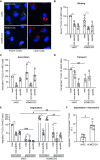
Figure 1
Figure 1
LDL is transcytosed through hAEC but degraded by hCMEC/D3. (A) HAEC and hCMEC/D3 were incubated with atto-655 LDL (50 μg/mL). After 3 h endothelial cells were fixed, counterstained with DAPI, and imaged using fluorescence microscopy. Representative images of n = 2 biological replicates, scale bar: 20 μm. Endothelial cells were incubated at 4 °C (binding (B)) or 37 °C (association (C)) for 1 h with 10 μg/mL of I125-LDL without (total) or with 40× excess of non-labeled LDL (unspecific). The specific LDL binding or association was calculated by subtracting the unspecific from the total binding or association, respectively. (D) Endothelial cells were grown on transwell inserts until confluence before adding 10 μg/mL of I125-LDL without (total) or with a 40× excess of LDL (unspecific) to the apical chamber. After 1 h, basolateral media were collected and radioactivity was measured using a gamma counter. Specific transport was calculated as described for the binding and association. (E) Endothelial cells were incubated with 10 μg/mL of I125-LDL without (total) or with a 40× excess of LDL (unspecific). After 4 h, media were collected, and cells were lysed in 0.2 mM NaOH. Degraded I125-LDL in the media was measured after TCA precipitation and compared to cell association. (F) The percentage of degradation per association was calculated by dividing the cpm of degradation by the sum of association + degradation ×100. Points in graphs represent individual experiments (biological replicates, n = 3–5), bars represent the mean and error bars ± SD: *, # p = 0.05, **, ## p = 0.01, and ***, ### p = 0.001.
Figure 2
Figure 2
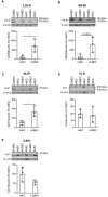
Brain and aortic endothelial cells express low-density lipoprotein receptor (LDLR), scavenger receptor (SR-BI), activin-like kinase (ALK1), clathrin heavy chain (CLH), and caveolin 1 (CAV1). hAEC (from three independent individuals) and hCMEC/D3 (from three successive passages) were grown until confluence before being lysed in RIPA buffer. Equal amounts of proteins (15–25 μg) were separated by SDS-PAGE. LDLR (A), SR-BI (B), ALK1 (C), CLH (D), and CAV1 (E) protein levels were measured by Western blotting and normalized to β-actin. Quantifications of band densitometry were performed using ImageJ. Points in graphs represent individual experiments (hAEC n = 3 individuals and hCMEC/D3 n = 3 successive cell passages), bars represent the mean and error bars ± SD, * p = 0.05.
Figure 3
Figure 3
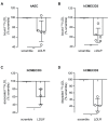
Knocking down LDLR reduces LDL binding, association, and degradation in hCMEC/D3. Seventy-two hours after silencing LDLR using siRNA, I125-LDL binding (4 °C) to hAEC (A) or hCMEC/D3 (B) was measured as described in Figure 1. To further investigate the interaction of LDL with hCMEC/D3, I125-LDL association (1 h at 37 °C) (C) and degradation (4 h at 37 °C) (D) were measured as above. Points in graphs represent individual experiments (biological replicates, n = 3–5), bars represent the mean and error bars ± SD, * p = 0.05 and ** p = 0.01.
Figure 4
Figure 4
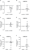
Inhibition of SR-BI reduces LDL binding to hAEC but not to hCMEC/D3. Seventy-two hours after silencing SCARB1 using siRNA, I125-LDL binding (4 °C) to hAEC (A) or hCMEC (B) was measured as above. I125-LDL association (37 °C) to hCMEC/D3 was measured as above 72 h after RNA interference against SCARB1 (C) or after treatment with an antibody blocking SR-BI (D). The role of SR-BI selective uptake was assessed by treating hCMEC/D3 with 1 μM BLT1. After 30 min, I125-LDL (E) or DiI-LDL (F) association were measured as above. Points in graphs represent individual experiments (biological replicates, n = 4–6), bars represent the mean and error bars ± SD, * p = 0.05.
Figure 5
Figure 5
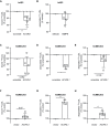
Knockdown and overexpression of ACVRL1 reduces and increases LDL degradation by hCMEC/D3, respectively. (A) Seventy-two hours after silencing ACVRL1 using siRNA, I125-LDL binding (4 °C) to hAEC was measured as above. (B) hAEC were incubated with 10 ng/mL of BMP-9 for 2 h before measuring I125-LDL association. (C) Seventy-two hours after silencing ACVRL1 using siRNA, I125-LDL binding to hCMEC/D3 was measured. To further investigate the interaction of LDL with hCMEC/D3, I125-LDL association (1 h at 37 °C) (D) and degradation (4 h at 37 °C) (E) were measured as above. After stable transfection of hCMEC/D3 with plasmid encoding for ALK1 and selection of the cells with the antibiotic G418, I125-LDL binding (F), association (G), and degradation (H) were measured as above. Points in graphs represent individual experiments (biological replicates, n = 3–5), bars represent the mean and error bars ± SD, * p = 0.05 and ** p = 0.01.
Figure 6
Figure 6
Knocking down AP2M1 but not caveolin reduces LDL association to hCMEC/D3. Seventy-two hours after knocking down AP2M1 (A) or CAV1 (B) using siRNA, I125-LDL association was measured as above. Points in graphs represent individual experiments (biological replicates, n = 3–4), bars represent the mean and error bars ± SD, * p = 0.05.

Similar articles

Cited by

References

    1. Jang E., Robert J., Rohrer L., von Eckardstein A., Lee W.L. Transendothelial Transport of Lipoproteins. Atherosclerosis. 2020;315:111–125. doi: 10.1016/j.atherosclerosis.2020.09.020. - DOI - PubMed
    1. Armstrong S.M., Sugiyama M.G., Fung K.Y.Y., Gao Y., Wang C., Levy A.S., Azizi P., Roufaiel M., Zhu S.N., Neculai D., et al. A Novel Assay Uncovers an Unexpected Role for SR-BI in LDL Transcytosis. Cardiovasc. Res. 2015;108:268–277. doi: 10.1093/cvr/cvv218. - DOI - PMC - PubMed
    1. Kraehling J.R., Chidlow J.H., Rajagopal C., Sugiyama M.G., Fowler J.W., Lee M.Y., Zhang X., Ramírez C.M., Park E.J., Tao B., et al. Genome-Wide RNAi Screen Reveals ALK1 Mediates LDL Uptake and Transcytosis in Endothelial Cells. Nat. Commun. 2016;7:13516. doi: 10.1038/ncomms13516. - DOI - PMC - PubMed
    1. Sun S.W., Zu X.Y., Tuo Q.H., Chen L.X., Lei X.Y., Li K., Tang C.K., Liao D.F. Caveolae and Caveolin-1 Mediate Endocytosis and Transcytosis of Oxidized Low Density Lipoprotein in Endothelial Cells. Acta Pharmacol. Sin. 2010;31:1336–1342. doi: 10.1038/aps.2010.87. - DOI - PMC - PubMed
    1. Goldstein J.L., Brown M.S. The LDL Receptor. Arterioscler. Thromb. Vasc. Biol. 2009;29:431–438. doi: 10.1161/ATVBAHA.108.179564. - DOI - PMC - PubMed

Publication types

MeSH terms

Grants and funding

This work was supported by a BrightFocus award (A2021037S) to J.R. and a Swiss National Science Foundation award (SNSF, 310030_185109) to A.V.E. G.P. was supported by a doctoral fellowship from the University of Zurich (Forschungskredit, grant no. FK-20-037).