. 2022 Oct 6;8(1):127.
doi: 10.1038/s41531-022-00394-9.

Cathepsin L-containing exosomes from α-synuclein-activated microglia induce neurotoxicity through the P2X7 receptor

Affiliations

Cathepsin L-containing exosomes from α-synuclein-activated microglia induce neurotoxicity through the P2X7 receptor

Tianfang Jiang et al. NPJ Parkinsons Dis. .

Abstract

Uncontrolled microglial activation is pivotal to the pathogenesis of Parkinson's disease (PD), which can secrete Cathepsin L (CTSL) to affect the survival of neurons in the PD patients; however, the precise mechanism has yet to be determined. We demonstrated for the first time that CTSL was mostly released by exosomes derived from α-Syn-activated microglia, resulting in neuronal damage and death. The elevation of CTSL activity was blocked by GW4869, suggesting a critical role for exosomes in mediating CTSL release. Furthermore, the P2X7R/PI3K/AKT signalling pathway was identified as the underlying molecular mechanism since specific antagonists of this signalling pathway, P2X7R knockdown and exosome release inhibitors significantly reduced the injury to cultured mouse cortical neurons. Our study suggests that increased extracellular release of CTSL from α-Syn-activated microglia through exosomes amplifies and aggravates of the neurotoxic effect of microglia, implying that CTSL may be involved in a fresh mechanism of PD pathogenesis, and serve as a potential biomarker and a target for PD drug development.


Conflict of interest statement

The authors declare no competing interests.

Figures

Fig. 1
Fig. 1. Extracellular α-Syn oligomers promote microglial CTSL and exosome release, and exosomes carry CTSL in α-Syn-activated microglia in a time-dependent manner.
a, b Western blot and quantitative analysis of CTSL release from microglia stimulated with extracellular WT or A53T α-Syn oligomer for up to 6 h (n = 4); *p < 0.05. c Representative TEM images of exosomes isolated from the supernatant of primary microglia. Scale bar: 200 nm. d, e Analysis of the particle number and size of microglia-derived exosomes from the different treatment groups by NTA and a Flow Nano Analyser (n = 4). f, g The results of Western blot analysis showing the presence of the exosomal markers CD63, TSG101 and Alix, and a high level of CTSL in exosomes from WT and A53T α-Syn oligomer-activated microglia (n = 4); ***p < 0.001. h LDH levels in the microglial culture medium were measured by ELISA after different treatments (n = 6). Ctrl = untreated control. Error bars represent s.e.m.
Fig. 2
Fig. 2. The main mechanism by which CTSL is released from α-Syn oligomers-activated is exosome-mediated release.
a Representative confocal microscopy images of immunofluorescence staining for CTSL and the specific exosome marker TSG101. When primary microglia were activated by treatment with WT or A53T α-Syn oligomer for up to 6 h, the colocalization of CTSL and TSG101 (indicated by arrows) increased over time (n = 3). Scale bar: 50 μm. b, c The cell supernatant and secreted exosomes were isolated from the same number of microglia, and the protein levels of CTSL in both the exosome-enriched and exosome-depleted fractions were assayed by western blotting (n = 4); *p < 0.05. d Measurement of CTSL activity in exosome-enriched lysates from untreated and α-Syn oligomers-treated microglia. Exosome lysates without the addition of substrate were used as background controls (n = 6); ***p < 0.001. e, f Exosomes were isolated from the supernatants of control microglial and WT and A53T α-Syn oligomer-treated microglia in the presence and absence of GW4869. CTSL, CD63, and TSG101 levels in exosome lysates were measured by western blotting (n = 4); *p < 0.05, **p < 0.01. Ctrl = untreated control; exo = exosome; GW5 = 5 μM GW4869; GW10 = 10 μM GW4869. Error bars represent s.e.m.
Fig. 3
Fig. 3. P2X7R is necessary for the release of CTSL-containing exosomes from α-Syn oligomers-treated microglia.
a Extracellular α-Syn oligomer interacts with P2X7R. Western blot analysis of α-Syn expression in microglia after treatment with WT or A53T α-Syn oligomer for up to 6 h. WT and A53T α-Syn oligomer were coimmunoprecipitated with P2X7R, with A53T α-Syn oligomer showing a stronger interaction. b Representative confocal microscopy images of immunofluorescence staining for α-Syn (red) and P2X7R (green) after treatment with A53T or WT α-Syn oligomer for up to 6 h to observe colocalization (indicated by arrows). Scale bar: 50 μm. c, d Western blot and quantitative analysis of CTSL release from the supernatant of microglia stimulated with BzATP (100 μM), an agonist of P2X7R, for up to 6 h (n = 4); *p < 0.05. e, f BzATP obviously induced CTSL release from α-Syn oligomers-activated microglia via exosomes, while pretreatment with BBG (1 μM), an antagonist of P2X7R, effectively reversed CTSL-containing exosome release (n = 4); *p < 0.05, **p < 0.01. g, h Western blot and quantitative analysis of P2X7R expression in microglia transfected with nonspecific (scramble siRNA) (20 nM) or P2X7R siRNA (20 nM) to confirm P2X7R knockdown. Densitometric analysis of the number of CTSL-containing exosomes released from microglia transfected with scramble or P2X7R siRNA after treatment with WT or A53T α-Syn oligomer for 6 h (n = 4); *p < 0.05, **p < 0.01, ***p < 0.001. Ctr = untreated control; Scr = scramble; siP2X7R = P2X7R siRNA. Error bars represent s.e.m.
Fig. 4
Fig. 4. P2X7R stimulates PI3K/AKT signalling in microglia in response to extracellular α-Syn oligomers, and α-Syn oligomers activate microglial CTSL-containing exosome release via the P2X7R/PI3K/AKT signalling pathway.
a, b p-AKT levels in microglia were increased after treatment with WT or A53T α-Syn oligomer, with A53T α-Syn oligomer causing a more significant increase in AKT phosphorylation. Inhibition of PI3K by LY294002 (50 μM) blocked α-Syn-induced phosphorylation of AKT (n = 4); *p < 0.05. c, d Exosomes were isolated from the supernatants of control and α-Syn oligomer-treated microglia in the presence or absence of LY294002. CTSL, CD63, and TSG101 levels in exosome lysates were quantitatively analysed by western blotting (n = 4); *p < 0.05. e, f p-AKT expression in microglia transfected with scramble or P2X7R siRNA after stimulation with WT or A53T α-Syn oligomer. Knockdown of P2X7R rescued the increase in p-AKT expression in BV2 cells in response to stimulation with WT or A53T α-Syn oligomer, implying that α-Syn oligomer-mediated stimulation of P2X7R is an upstream event that activates PI3K/AKT signalling and ultimately CTSL-containing exosome release (n = 4); *p < 0.05. Ctrl = untreated control; LY = LY294002; Scr = scramble; siP2X7R = P2X7R siRNA. Error bars represent s.e.m.
Fig. 5
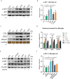
Fig. 5. CTSL-containing exosomes released from activated microglia promote apoptosis of neurons.
a, b Primary cortical neurons were treated with exosomes isolated from microglia treated with or without WT or A53T α-Syn oligomer. Western blot and quantitative analysis of cleaved-caspase-3 expression in neurons treated for 24 h with exosomes from untreated microglia or microglia treated with LPS or WT or A53T α-Syn oligomer (n = 3); ***p < 0.001. c, d The level neuronal apoptosis was determined after co-culture with exosomes from microglia pretreated with or without GW4869 (n = 3); *p < 0.05, **p < 0.01, ***p < 0.001. e, f The level of apoptosis of neurons cocultured with exosomes from microglia treated with or without iCL (n = 3); **p < 0.01, ***p < 0.001. g Western blot and quantitative analysis of CTSL expression in microglia transfected with nonspecific (scramble siRNA) (20 nM) or P2X7R siRNA (20 nM) to confirm CTSL knockdown. Exosomes secreted by α-Syn oligomers-activated CTSL knockdown microglia induce less neuronal death (n = 3). h Densitometric analysis of the number of CTSL-containing exosomes released from microglia transfected with scramble or CTSL siRNA after treatment with WT or A53T α-Syn oligomer for 6 h (n = 3); *p < 0.05, **p < 0.01, ***p < 0.001. The amount of exosomes used in each group was secreted by approximately 3*107 microglia cells. Ctrl = untreated control; Scr = scramble; siCTSL = CTSL siRNA; EC = exosomes from untreated microglia (the control group); EL = exosomes from LPS-treated microglia; EW = exosomes from WT α-Syn oligomer-treated microglia; EA = exosomes from A53T α-Syn oligomer-treated microglia; ED = exosomes from DMSO-treated microglia; EiCL = exosomes from iCL-treated microglia; GW5 = 5 μM GW4869; GW10 = 10 μM GW4869; EsiCTSL = exosomes from CTSL knockdown microglia. Error bars represent s.e.m.
Fig. 6
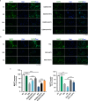
Fig. 6. Representative immunofluorescence images of neurons labelled with MAP2 illustrating the neurotoxic potential of CTSL-containing exosomes.
a, b Treatment with GW4869 or the CTSL inhibitor iCL (20 μM) prior to exosome isolation rescued CTSL-containing exosome-induced neurotoxicity in a dose-dependent manner in each experimental group, as determined by MAP2 immunostaining, suggesting a direct neurotoxic role for CTSL released from α-Syn oligomers-treated microglia. Scale bar: 50 μm. c Quantification of the percentage of MAP2+ neurons for the experiments depicted in panels a (up) and b (down). Results are representative of three fluorescent images of each group from three independent experiments. The amount of exosomes used in each group was secreted by approximately 104 microglia cells. EC = exosomes from untreated microglia (the control group); EW = exosomes from WT α-Syn oligomer-treated microglia; EA = exosomes from A53T α-Syn oligomer-treated microglia; ED = exosomes from DMSO-treated microglia; EiCL = exosomes from iCL-treated microglia; GW5 = 5 μM GW4869; GW10 = 10 μM GW4869. Error bars represent s.e.m.
Fig. 7
Fig. 7. Secretion of exosomes by microglia is responsible for the transmission of CTSL to neurons.
a Exosomes isolated from the same volume of culture medium from microglia exposed to different experimental conditions were labelled with the dye PKH67. These exosomes were then added to recipient neurons and incubated for 1 day. The neurons were then fixed with 4% PFA (n = 3). Exosomes secreted by α-Syn oligomers-treated microglia were endocytosed by neurons (spot-shaped, indicated by arrows). The amount of exosomes used in each group was secreted by approximately 104 microglia cells. Scale bar: 50 μm. b, c The level of CTSL, CD63 and TSG101 in neurons cocultured with exosomes from microglia pretreated with or without GW4869 was measured by western blotting and quantitatively analysed (n = 3); *p < 0.05, **p < 0.01. GW4869 treatment reduced exosome internalisation by neurons, resulting in lowers CTSL expression. d, e Pretreatment with the CTSL inhibitor iCL decreased the expression of CTSL in microglia, as determined by western blotting and quantitative analysis (n = 3); *p < 0.05, **p < 0.01, ***p < 0.001. f, g Due to the inhibitory effect of iCL, less CTSL was released by α-Syn-activated microglia and internalised by neurons (n = 3); *p < 0.05. h, i CTSL, CD63, and TSG101 expression in exosome lysates from microglia was measured by western blotting. Administration of iCL prior to activation with α-Syn did not affect the reduction in exosome release from microglia, which once again proves that CTSL, not exosomes, is responsible for neuronal damage (n = 3); ***p < 0.001. The amount of exosomes used in each group was secreted by approximately 3*107 microglia cells. EC = exosomes from untreated microglia (the control group); EW = exosomes from WT α-Syn-treated microglia; EA = exosomes from A53T α-Syn-treated microglia; ED = exosomes from DMSO-treated microglia; EiCL = exosomes from iCL-treated microglia; Ctrl = untreated control; GW5 = 5 μM GW4869; GW10 = 10 μM GW4869. Error bars represent s.e.m.

Similar articles

Cited by

References

    1. Blaylock RL. Parkinson’s disease: Microglial/macrophage-induced immunoexcitotoxicity as a central mechanism of neurodegeneration. Surg. Neurol. Int. 2017;8:65. doi: 10.4103/sni.sni_441_16. - DOI - PMC - PubMed
    1. Bartels T, De Schepper S, Hong S. Microglia modulate neurodegeneration in Alzheimer’s and Parkinson’s diseases. Science. 2020;370:66–69. doi: 10.1126/science.abb8587. - DOI - PubMed
    1. Lannes N, Eppler E, Etemad S, Yotovski P, Filgueira L. Microglia at center stage: a comprehensive review about the versatile and unique residential macrophages of the central nervous system. Oncotarget. 2017;8:114393–114413. doi: 10.18632/oncotarget.23106. - DOI - PMC - PubMed
    1. Jiang T, et al. P2X7 receptor is critical in alpha-synuclein–mediated microglial NADPH oxidase activation. Neurobiol. Aging. 2015;36:2304–2318. doi: 10.1016/j.neurobiolaging.2015.03.015. - DOI - PubMed
    1. Tamhane T, et al. Nuclear cathepsin L activity is required for cell cycle progression of colorectal carcinoma cells. Biochimie. 2016;122:208–218. doi: 10.1016/j.biochi.2015.09.003. - DOI - PubMed