Review
. 2022 Oct 5;7(1):353.
doi: 10.1038/s41392-022-01200-x.

Signaling pathways and targeted therapies in lung squamous cell carcinoma: mechanisms and clinical trials

Affiliations
Review

Signaling pathways and targeted therapies in lung squamous cell carcinoma: mechanisms and clinical trials

Zhenyi Niu et al. Signal Transduct Target Ther. .

Abstract

Lung cancer is the leading cause of cancer-related death across the world. Unlike lung adenocarcinoma, patients with lung squamous cell carcinoma (LSCC) have not benefitted from targeted therapies. Although immunotherapy has significantly improved cancer patients' outcomes, the relatively low response rate and severe adverse events hinder the clinical application of this promising treatment in LSCC. Therefore, it is of vital importance to have a better understanding of the mechanisms underlying the pathogenesis of LSCC as well as the inner connection among different signaling pathways, which will surely provide opportunities for more effective therapeutic interventions for LSCC. In this review, new insights were given about classical signaling pathways which have been proved in other cancer types but not in LSCC, including PI3K signaling pathway, VEGF/VEGFR signaling, and CDK4/6 pathway. Other signaling pathways which may have therapeutic potentials in LSCC were also discussed, including the FGFR1 pathway, EGFR pathway, and KEAP1/NRF2 pathway. Next, chromosome 3q, which harbors two key squamous differentiation markers SOX2 and TP63 is discussed as well as its related potential therapeutic targets. We also provided some progress of LSCC in epigenetic therapies and immune checkpoints blockade (ICB) therapies. Subsequently, we outlined some combination strategies of ICB therapies and other targeted therapies. Finally, prospects and challenges were given related to the exploration and application of novel therapeutic strategies for LSCC.


Conflict of interest statement

The authors declare no competing interests.

Figures

Fig. 1
Fig. 1
Timeline illustrating the evolving treatment landscapes and research history of LSCC. Timeline highlights some of the pivotal clinical studies and discoveries that might change or have changed the management of LSCC. 1 L first line, 2 L second line, ICB therapy immune-checkpoint blockade therapy, OS overall survival, TCGA The Cancer Genome Atlas
Fig. 2
Fig. 2
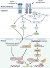
Mutated driver genes and downstream signal pathways in LSCC. FGFR1 amplification is observed in 20% of LSCC patients. FGFR1 amplifications can lead to overexpression of wild-type FGFR1 proteins on the cell membrane, resulting in increased sensitivity to FGF and the promotion of tumor growth via increased activation of four key downstream signaling pathways: PLCγ, PI3K-AKT, RAS-MAPK, and STAT (green). Although mutations in EGFR gene are relatively rare in LSCC, in certain cases of LSCC these mutations are responsible for constitutive ligand-independent receptor activation and downstream signaling, promoting cell survival and proliferation. Mitogenic signaling, including activation of upstream PI3K and MAPK signaling, could drive cyclin D upregulation, which leads to CDK4/6 activation. The phosphorylation of RB by cyclin D-CDK4/6 complexes and cyclin E-CDK2 complexes releases E2F transcriptional factors to activate genes required for G1-S transition. The CDKN2A gene encodes p16INK4A and p14ARF, which regulate cell cycle by inhibiting CDK4/6 and CDK2, respectively. In LSCC, the inactivated CDKN2A caused by genomic alterations may upregulate this pathway. DAG diacylglycerol, E2F E2 family, FRS2 FGFR substrate 2, GRB2 growth factor receptor-bound 2, IP3 inositol triphosphate, MDM2 murine double minute 2, P phosphorylation, PIP2 phosphatidylinositol-4,5-biphosphate, PKC protein kinase C, PLCγ phospholipase Cγ, PTEN phosphatase and tensin homolog, STAT signal transducer and activator of transcription
Fig. 3
Fig. 3
Physiologic activation and regulation of NRF2 and metabolic reprogramming by NRF2 in LSCC cells. a In unstressed conditions, KEAP1 forms a ubiquitin E3 ligase complex with CULLIN3 (CUL3) and binds with NFR2 via the DLG and ETGE motifs in the Neh2 domain of NRF2 in the cytoplasm. NRF2 is then polyubiquitinated and degraded through the proteasome system after its synthesis. When cells are exposed to electrophiles or ROS, KEAP1 is modified and the KEAP1-CUL3 ubiquitin E3 ligase activity declines, which stabilizes NRF2. Stabilized and accumulated NRF2 translocates to the nucleus and functions as a transcriptional factor. NRF2 is also regulated through a KEAP1-independent mechanism in which GSK3 plays an important role. NRF2 is phosphorylated by GSK3 and then recognized by β-TrCP. By contrast, the Neh6 domain of NRF2 serves as the degron exploited in this β-TrCP-CUL1-dependent degradation of NRF2. Following its ubiquitination by the β-TrCP-CUL1 E3 ubiquitin ligase complex, NRF2 is degraded by the proteasome. b LSCC cells displayed a dual reliance on glucose and glutamine metabolism. Activation of NRF2 increases the synthesis of GSH from intracellular glutamate, cysteine, and glycine. GLS1 catalyzes the transformation of glutamine to glutamate. Cystine is imported by the xc antiporter system (xCT). Serine and glycine are synthesized via NRF2-dependent processes. Under chronic mTOR inhibition which suppresses glycolysis, LSCC cells could upregulate glutaminolysis through the GSK3 signaling pathway which developed acquired resistance to mTOR inhibition. β-TrCP β-transducin repeat-containing protein genes, GLS1 glutaminase 1, GSH glutathione, PDK phosphoinositide-dependent kinase, ROS reactive oxygen species, TCA cycle tricarboxylic acid cycle
Fig. 4
Fig. 4
Schematic diagram of the different roles for epigenetic therapeutic targets in LSCC. a A recent study suggested that NSD3, the neighboring gene of FGFR1, rather than FGFR1, was the critical driver oncogene within this recurrent focal amplicon of 8p11-12 genomic region. The amplification of NSD3 leads to increased NSD3 expression, thus increasing the synthesis of H3K36me2. Less common than the amplification of 8p11-12 and NSD3 expression, the GOF variant NSD3 was also present in LSCC. These two works together to increase H3K36me2, stimulating transcription of oncogenic targets, including mTOR pathways and MYC-associated pathways. This process rendered the tumor NSD3-addicted, which could be inhibited by BETi. b SOX2 and BCL11A are both identified as LSCC oncogenes. The BCL11A-SOX2 transcriptional program is crucial for the maintenance of a squamous phenotype. SETD8 is a monomethyltransferase, whose gene is regulated by SOX2 and BCL11A. The inhibition of SETD8 selectively limits LSCC tumor growth. c LSD1 could promote tumorigenesis in two different ways. The first way is demethylase-dependent. In SOX2-expressing tumor cells, LSD1 inhibition will induce increased H3K9me1/me2. The repressive H3K9 methylations act on the SOX2 gene, leading to SOX2 downregulation, reduced oncogenic potential, and increased cellular differentiation. The second way is demethylase-independent. In cells with a low level of LSD1, FBXW7 forms a dimer, which promotes ubiquitylation for proteasomal degradation of oncoprotein substrates, thus suppressing cell outgrowth. In cancer cells with overexpressed LSD1, the FBXW7 dimerization is blocked by LSD1 binding to FBXW7 in a demethylase-independent manner. FBXW7 self-ubiquitylation will then be triggered, followed by degradation by proteasome as well as lysosome in a p62-dependent pathway. d EZH2 is an enzymatic subunit of PRC2, which also includes EED, SUZ12, and RBBP4/7. The SET domain of EZH2 is responsible for the catalyzes the mono-, di-, and trimethylation of H3K27 from the universal methyl donor SAM, after which SAM becomes SAH. EZH2 also has noncanonical functions with its hidden TAD. The EZH2 TAD directly interacts with cMyc and other activators, including p300 and SWI/SNF. GOF gain-of-function, PRC2 polycomb repressive complex 2, SAH S-adenosyl-l-homocysteine, SAM S-adenosyl-l-methionine, TAD transactivation domain
Fig. 5
Fig. 5
Roles of Fcγ receptors in anti-CTLA-4 function. Selective deletion of Tregs in the tumor microenvironment results in tumor immunity (left). Expressing higher levels of CTLA-4 than effector T cells, intratumoral Tregs are selectively depleted through ADCP by macrophages and/or ADCC by NK cells. In T-effector cells, T-cell activity is enhanced by the recognition of MHC-Ag by the TCR in the presence of an anti-CTLA-4 antibody that had co-engaged with FcγR on APCs (right). ADCC antibody-dependent cellular cytotoxicity, ADCP antibody-dependent cellular phagocytosis, APC antigen-presenting cells, MHC-Ag major histocompatibility complex-antigen peptide complexes major histocompatibility complex-antigen peptide complexes, NK cells natural killer cells, TCR T-cell receptor
Fig. 6
Fig. 6
Impact of oncogenic signaling on tumor immune response. a Loss of PTEN protein function and improper PI3K activation inhibit efficient LC3 lipidation, which further promote resistance to T-cell-mediated killing by inhibiting autophagy. PTEN loss could also induce expression of immunosuppressive cytokines, including CCL12 and VEGF. b CDK4/6 inhibition enhances T-cell activation through the derepression of NFAT family proteins and their target genes, which encodes critical regulators of T-cell function. CDK4/6 inhibition could also induce Rb-mediated G1-arrest and promote the phenotypic and functional acquisition of immunologic T-cell memory. Besides, the PD-L1 protein stability is regulated by the CDK4-SPOP-FZR1 signaling pathway. Physiologically, PD-L1 protein stability is negatively regulated through phosphorylating its upstream physiological E3 ligase SPOP. This phosphorylation promotes SPOP binding to 14-3-3γ, which subsequently disrupts FZR1-mediated destruction of SPOP. The inhibition of CDK4/6 inhibits the phosphorylation of SPOP, thus promoting its degradation by FZR1, thus increasing PD-L1 protein levels. c Tumor-derived VEGF limits NF-κB activation in immature DCs, which in turn leads to defective functional maturation of DCs and insufficient induction of tumor immunity. VEGF could also impact the endothelial cells expression of immunological molecules. It decreases the expression of VCAM-1, which is important for the antitumor T cells adhesion and infiltration into tumors. Besides, VEGF also increases the expression of FAS ligand on endothelial cells, triggering apoptosis of T cells. VEGF also promotes the expansion of immune suppressive MDSCs, which further promotes the recruitment of Tregs. d EZH2 inhibition increases the production of CXCL9 and CXCL10, which are attractant cytokines promoting trafficking of T cells to tumor. Besides, EZH2 inhibition could selectively target intratumeral Tregs and reduce its immunosuppressive capacity. e In tumor cells, the ablation of LSD1 in cancer cells increases repetitive element expression, including ERVs, and decreases expression of RISC components. This leads to dsRNA stress and activation of type 1 interferon, which stimulates antitumor T-cell infiltration. In addition, inhibiting LSD1 in CD8+ T cells unleashes the transcription program mediated by TCF1, which is critical for the maintenance of the progenitor subset of intratumoral CD8+ T cells for persistent tumor control. dsRNA double-stranded RNA, ERVs endogenous retroviral elements, MDSCs myeloid-derived suppressor cells, RISC RNA-induced silencing complex, TCF1 T-cell factor 1, VCAM-1 vascular cell adhesion molecule-1

Similar articles

Cited by

References

    1. Siegel RL, Miller KD, Fuchs HE, Jemal A. Cancer statistics, 2022. CA Cancer J. Clin. 2022;72:7–33. doi: 10.3322/caac.21708. - DOI - PubMed
    1. Sung H, et al. Global cancer statistics 2020: GLOBOCAN estimates of incidence and mortality worldwide for 36 cancers in 185 countries. CA Cancer J. Clin. 2021;71:209–249. doi: 10.3322/caac.21660. - DOI - PubMed
    1. Herbst RS, Morgensztern D, Boshoff C. The biology and management of non-small cell lung cancer. Nature. 2018;553:446–454. doi: 10.1038/nature25183. - DOI - PubMed
    1. Heist RS, Sequist LV, Engelman JA. Genetic changes in squamous cell lung cancer: a review. J. Thorac. Oncol. 2012;7:924–933. doi: 10.1097/JTO.0b013e31824cc334. - DOI - PMC - PubMed
    1. Society, A. C. Cancer facts & figures 2022. https://www.cancer.org/content/dam/cancer-org/research/cancer-facts-and-... (2022).

Publication types

MeSH terms

Substances