. 2022 Oct 4;13(10):848.
doi: 10.1038/s41419-022-05299-2.

Upregulation of CRABP2 by TET1-mediated DNA hydroxymethylation attenuates mitochondrial apoptosis and promotes oxaliplatin resistance in gastric cancer

Affiliations

Upregulation of CRABP2 by TET1-mediated DNA hydroxymethylation attenuates mitochondrial apoptosis and promotes oxaliplatin resistance in gastric cancer

Xiaolong Tang et al. Cell Death Dis. .

Abstract

Oxaliplatin is the main chemotherapy drug for gastric cancer (GC), but quite a few patients are resistant to oxaliplatin, which contributes to the poor prognosis of GC patients. There is therefore an urgent need to identify potential targets for reversing chemotherapy resistance in GC patients. In this study, we analyzed the tumor samples of GC patients who received neoadjuvant chemotherapy based on oxaliplatin through quantitative proteomics and identified the potential chemoresistance-related protein cellular retinoic acid binding protein 2 (CRABP2). CRABP2 was significantly upregulated in the tumor tissues of chemoresistant GC patients and was closely related to prognosis. The results of cell function experiments showed that CRABP2 can promote the oxaliplatin resistance of GC cells in vitro. Coimmunoprecipitation and GST pulldown assays showed that CRAPB2 expedited the binding of BAX and PARKIN in GC cells and facilitated the ubiquitination-mediated degradation of BAX. Furthermore, both the in vitro assay and cell-derived xenograft (CDX) in vivo model verified that CRABP2 promoted oxaliplatin resistance by inhibiting BAX-dependent cell apoptosis. Further experiments proved that the abnormally high expression of CRABP2 in oxaliplatin-resistant GC cells was affected by TET1-mediated DNA hydroxymethylation. The patient-derived xenograft (PDX) model suggested that interference with CRABP2 reversed oxaliplatin resistance in GC in vivo. In conclusion, the results of our study show that CRABP2 was a key molecule in oxaliplatin resistance regulation and could be a new target for reversing the chemoresistance of GC.


Conflict of interest statement

The authors declare no competing interests.

Figures
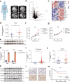
Fig. 1
Fig. 1. The expression of CRABP2 was upregulated in chemoresistant GC patients and predicted prognosis.
a Typical image examples of CR and CS gastric cancer patients who received NAC. The volcano map (b) and heatmap (c) of the most upregulated/downregulated differentially expressed proteins in CR and CS tumor tissues. The qRT–PCR (d, n = 22) and western blotting (e, n = 4) results showed the expression of CRABP2 in tumor tissues of CR and CS patients. The CCK-8 results of AGS cells treated with OXA (f) or fluorouracil (g) after knockdown/overexpression of CRABP2. h The relative expression of CRABP2 in parental and OXA-resistant GC cells by qRT–PCR. i The expression of CRABP2 from the TCGA database (tumor = 335; normal = 26). Mann–Whitney U test (Wilcoxon rank sum test). The outlier values (median±2 IQR) and extreme values (median ± 3.5 IQR) were excluded. IQR: interquartile range. The expression of CRABP2 in tumor tissues and adjacent normal tissues of GC patients by qRT–PCR (j, n = 44) and western blotting (k, n = 12). l Typical IHC images of CRABP2-positive and CRABP2-negative tumor tissues. m The correlation curves between CRABP2 expression and OS of GC patients (n = 488). (CR chemotherapy resistance, CS chemotherapy sensitivity, OXA oxaliplatin, C tumor tissue, N normal tissues, IHC immunohistochemistry, OS overall survival). *P < 0.05, **P < 0.01, ***P < 0.001.
Fig. 2
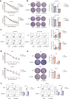
Fig. 2. CRABP2 promoted the oxaliplatin resistance of GC cells.
a After knocking down CRABP2, the viability of AGS-OXA and HGC-27-OXA cells was determined under different concentrations of OXA. b After knocking down CRABP2, the colony forming ability of AGS-OXA and HGC-27-OXA cells was determined in the absence or presence of OXA (concentration: 2.0 μM). c The cell apoptosis results of AGS-OXA and HGC-27-OXA cells after knocking down CRABP2 in the presence or absence of OXA (concentration: 2.0 μM) by flow cytometry. d In the presence of different concentrations of OXA, AGS and HGC-27 cells overexpressing CRABP2 were determined by cell viability assay. e In the presence or absence of OXA (concentration: AGS 0.2 μM, HGC-27 0.4 μM), AGS and HGC-27 cells overexpressing CRABP2 were determined by the colony forming assay. f In the absence or presence of OXA (concentration: AGS 0.2 μM, HGC-27 0.4 μM), the cell apoptosis results of AGS and HGC-27 cells overexpressing CRABP2 were determined by flow cytometry. All experiments were performed in three replicates. The data are presented as the mean ± SD. *P < 0.05, **P < 0.01, ***P < 0.001.
Fig. 3
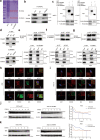
Fig. 3. CRABP2 expedited the binding ability of BAX and PARKIN in GC.
a Total proteins from Flag-CRABP2 plasmid-transfected AGS cells were separated via SDS–PAGE. PARKIN and BAX were identified by LC/LC–MS in the CRABP2 protein complex. b Mutual interactions of BAX/PARKIN and Flag-CRABP2 were verified by the Co-IP assay. c The BAX and PARKIN proteins were pulled down by GST-CRABP2 fusion protein-bound beads by SDS–PAGE analysis. d The interaction between BAX and CRABP2 and between PARKIN and CRABP2 was verified by co-IP assay and western blotting. e After interference with CRABP2 in AGS cells, the binding ability of BAX to PARKIN decreased. f After BAX interference in AGS cells, the binding ability of CRABP2 to PARKIN was not affected. g After interference with PARKIN in AGS cells, the binding ability of CRABP2 to PARKIN was not affected. The immunofluorescence assay showed the subcellular localizations (red frame) of CRABP2/BAX/PARKIN in AGS (h) and HGC-27 (i) cells. AGS cells overexpressing (j) or knocking down (k) CRABP2 were treated with cycloheximide (CHX) for 0, 2, 4, 6, 8, or 10 h. The expression of Bax was detected by western blotting. All experiments were performed in three replicates. *P < 0.05, **P < 0.01, ***P < 0.001. (IB immunoblotting, IP immunoprecipitation, WCL whole cell lysates).
Fig. 4
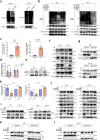
Fig. 4. CRABP2 promoted chemoresistance by regulating ubiquitination degradation of Bax.
a After adding MG-132 to AGS cells with knockdown or overexpression of CRABP2, Co-IP of Bax was performed, and ubiquitin was detected by western blotting. b GC cells with CRABP2 knockdown or overexpression were transfected with HA-labeled ubiquitinated plasmids of different sites, and Co-IP experiments of HA were performed. After adding OXA to AGS/HGC-27 cells, the activities of caspase 9 (c) and caspase 12 (d) were detected. e After adding OXA to AGS/HGC-27 cells, the expression of CC3 was detected. f After adding OXA to AGS/HGC-27 cells, the expression of DNA damage/repair-related proteins was examined by western blotting. g After AGS/HGC-27 cells were treated with OXA, the cytoplasm and mitochondria were separated, and the expression of BAX was examined. h The activities of caspase 9 were detected after CRABP2 knockdown/overexpression in AGS/HGC-27 cells. i The expression of CC3 was examined after CRABP2 knockdown/overexpression and OXA added to AGS/HGC-27 cells. j After adding OXA to AGS/HGC-27 cells and knocking down/overexpressing CRABP2, the expression of DNA damage/repair-related proteins was examined by western blotting. k, l After AGS/HGC-27 cells with CRABP2 knockdown or overexpression were treated with OXA, the cytoplasm and mitochondria were separated, and the expression of BAX was examined. (OXA oxaliplatin, CC3 cleaved caspase 3, TOMM40 translocase of outer mitochondrial membrane 40).
Fig. 5
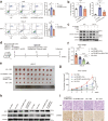
Fig. 5. CRABP2 promoted oxaliplatin resistance through the BAX-dependent cell apoptosis pathway.
a After knocking down CRABP2/ BAX and adding OXA (concentration: AGS 0.2 μM, HGC-27 0.4 μM), the percentage of apoptotic cells was examined by flow cytometry. b After knocking down CRABP2/ BAX and adding OXA (concentration: AGS 0.2 μM, HGC-27 0.4 μM), the activity of cleaved caspase 9 was examined. c After knocking down CRABP2/ BAX and adding OXA (concentration: AGS 0.2 μM, HGC-27 0.4 μM), the expression of cleaved caspase 3/BAX/CRABP2 was examined by western blotting. d Schematic diagram of the experimental procedures of the cell-derived xenograft model in nude mice. e Photo showing tumor formation in the four groups of nude mice. Tumor weight (f) and tumor volumes (g) of four groups of nude mice. h The expression of CRABP2 and BAX in tumors from four groups of nude mice by western blotting. i HE and CRABP2 IHC staining of tumors in four groups of nude mice. All experiments were performed in three replicates. *P < 0.05, **P < 0.01, ***P < 0.001.
Fig. 6
Fig. 6. TET1-mediated DNA hydroxymethylation expedited the expression of CRABP2.
a After adding 5AZ, Bobcat339, DZNEP, TSA, or SAHA to AGS and HGC-27 cells, the mRNA expression changes in CRABP2 were detected. The methylation (b) and hydroxymethylation (c) levels of OXA-resistant GC cells and parental GC cells were examined. d The expression and activity of DNA methylase, demethylase, and hydroxymethylase from OXA-resistant and parental GC cells were examined by western blotting. e The activity of hydroxymethylase TET1 was detected in OXA-resistant GC cells and parental GC cells. f Schematic diagram of the predicted and designed methylation sites in the promoter region of CRABP2. g ChIP experiments using a TET1 antibody were performed in OXA-resistant GC cells. h Hydroxymethylated ChIP assays were performed in OXA-resistant GC cells. After knocking down or overexpressing TET1 in OXA-resistant GC cells, the expression of CRABP2 was examined by qRT–PCR (i) and western blotting (j). *P < 0.05, **P < 0.01, ***P < 0.001, ns not significant.
Fig. 7
Fig. 7. Interference with CRABP2 reversed oxaliplatin resistance in GC in vivo.
a Schematic diagram of the patient-derived xenograft model of GC. b Photos of different tumor formations in the two groups of NOD-SCID mice. Tumor weight (c) and tumor volume (d) measurement in the two groups of mice. e Expression of CRABP2 and BAX in tumors from the two groups of mice, as determined by western blotting. f HE staining and CRABP2 staining of tumor slides from the two groups of mice. g Under normal physiological conditions, the synthesis and degradation of Bax in the cytoplasm are in equilibrium. When cells are treated with oxaliplatin, Bax integrates into the outer mitochondrial membrane, leading to the release of cytochrome C and cell apoptosis. In chemoresistant GC cells, in which the expression of CRABP2 in the cell is at a high level, CRABP2 can promote the ubiquitination degradation of Bax by combining Bax and Parkin, ultimately allowing the cells to survive. *P < 0.05, **P < 0.01, ***P < 0.001.

Similar articles

Cited by

References

    1. Siegel RL, Miller KD, Jemal A. Cancer statistics, 2019. CA Cancer J Clin. 2019;69:7–34. doi: 10.3322/caac.21551. - DOI - PubMed
    1. Zeng H, Chen W, Zheng R, Zhang S, Ji JS, Zou X, et al. Changing cancer survival in China during 2003-15: a pooled analysis of 17 population-based cancer registries. Lancet Glob Health. 2018;6:e555–e67.. doi: 10.1016/S2214-109X(18)30127-X. - DOI - PubMed
    1. Noh SH, Park SR, Yang HK, Chung HC, Chung IJ, Kim SW, et al. Adjuvant capecitabine plus oxaliplatin for gastric cancer after D2 gastrectomy (CLASSIC): 5-year follow-up of an open-label, randomised phase 3 trial. Lancet Oncol. 2014;15:1389–96. doi: 10.1016/S1470-2045(14)70473-5. - DOI - PubMed
    1. Martinez-Balibrea E, Martinez-Cardus A, Gines A, Ruiz de Porras V, Moutinho C, Layos L, et al. Tumor-Related Molecular Mechanisms of Oxaliplatin Resistance. Mol Cancer Ther. 2015;14:1767–76. doi: 10.1158/1535-7163.MCT-14-0636. - DOI - PubMed
    1. Joshi SS, Badgwell BD. Current treatment and recent progress in gastric cancer. CA Cancer J Clin. 2021;71:264–79.. doi: 10.3322/caac.21657. - DOI - PMC - PubMed

Publication types

MeSH terms