

Breast cancers co-opt normal mechanisms of tolerance to promote immune evasion and metastasis
- PMID: 36189970
- PMCID: PMC9662806
- DOI: 10.1152/ajpcell.00189.2022
Breast cancers co-opt normal mechanisms of tolerance to promote immune evasion and metastasis
Abstract
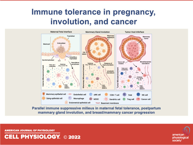
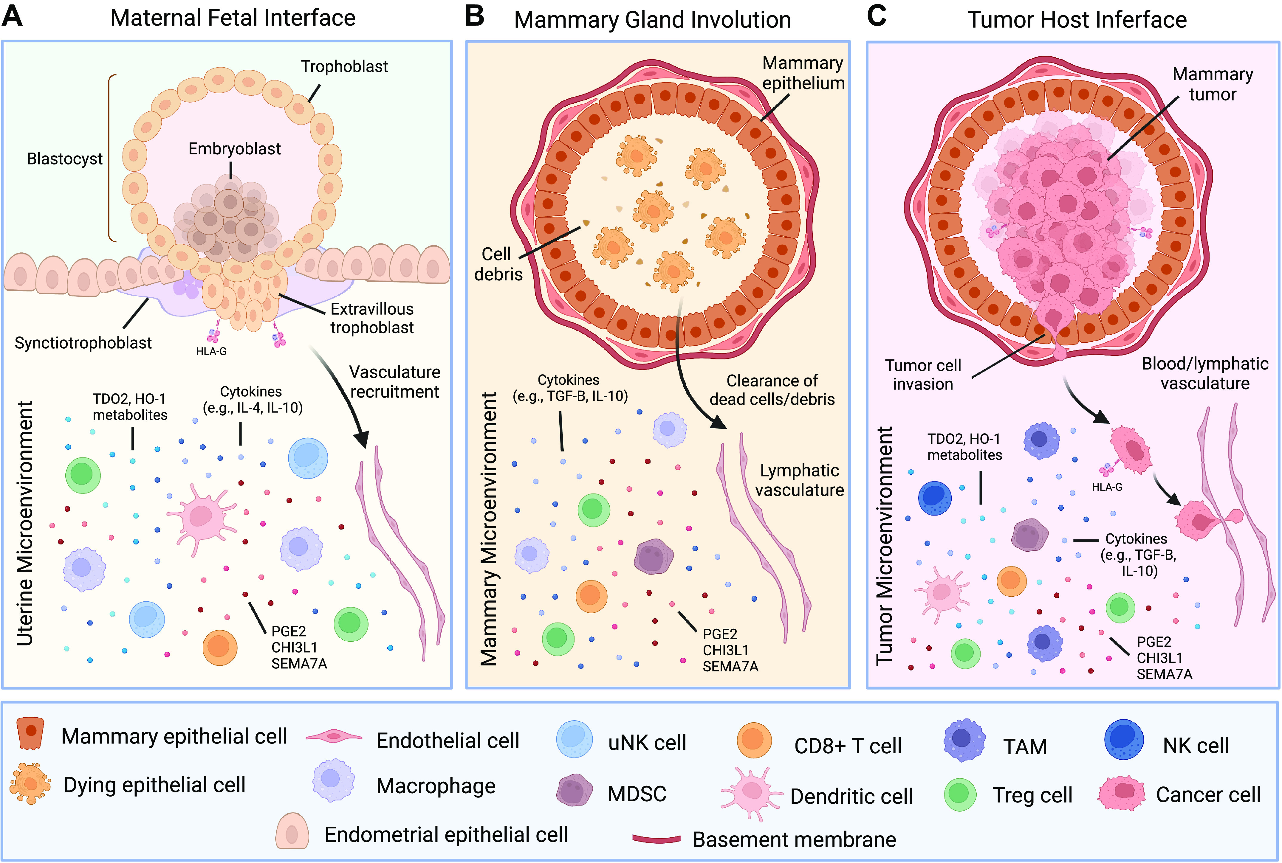
Normal developmental processes, such as those seen during embryonic development and postpartum mammary gland involution, can be reactivated by cancer cells to promote immune suppression, tumor growth, and metastatic spread. In mammalian embryos, paternal-derived antigens are at risk of being recognized as foreign by the maternal immune system. Suppression of the maternal immune response toward the fetus, which is mediated in part by the trophoblast, is critical to ensure embryonic survival and development. The postpartum mammary microenvironment also exhibits immunosuppressive mechanisms accompanying the massive cell death and tissue remodeling that occurs during mammary gland involution. These normal immunosuppressive mechanisms are paralleled during malignant transformation, where tumors can develop neoantigens that may be recognized as foreign by the immune system. To circumvent this, tumors can dedifferentiate and co-opt immune-suppressive mechanisms normally utilized during fetal tolerance and postpartum mammary involution. In this review, we discuss those similarities and how they can inform our understanding of cancer progression and metastasis.
Keywords: cancer; inflammation; involution; macrophages; pregnancy.
Conflict of interest statement
No conflicts of interest, financial or otherwise, are declared by the authors.
Figures
Similar articles
-
Wound healing-like immune program facilitates postpartum mammary gland involution and tumor progression.Int J Cancer. 2015 Apr 15;136(8):1803-13. doi: 10.1002/ijc.29181. Epub 2014 Sep 15. Int J Cancer. 2015. PMID: 25187059 Free PMC article.
-
Semaphorin 7A Promotes Macrophage-Mediated Lymphatic Remodeling during Postpartum Mammary Gland Involution and in Breast Cancer.Cancer Res. 2018 Nov 15;78(22):6473-6485. doi: 10.1158/0008-5472.CAN-18-1642. Epub 2018 Sep 25. Cancer Res. 2018. PMID: 30254150 Free PMC article.
-
Postpartum breast involution reveals regression of secretory lobules mediated by tissue-remodeling.Breast Cancer Res. 2014 Mar 28;16(2):R31. doi: 10.1186/bcr3633. Breast Cancer Res. 2014. PMID: 24678808 Free PMC article.
-
Macphatics and PoEMs in Postpartum Mammary Development and Tumor Progression.J Mammary Gland Biol Neoplasia. 2020 Jun;25(2):103-113. doi: 10.1007/s10911-020-09451-6. Epub 2020 Jun 13. J Mammary Gland Biol Neoplasia. 2020. PMID: 32535810 Free PMC article. Review.
-
A mouse mammary gland involution mRNA signature identifies biological pathways potentially associated with breast cancer metastasis.J Mammary Gland Biol Neoplasia. 2009 Jun;14(2):99-116. doi: 10.1007/s10911-009-9120-1. Epub 2009 Apr 30. J Mammary Gland Biol Neoplasia. 2009. PMID: 19408105 Review.
Cited by
-
Proteomic characterization identifies clinically relevant subgroups of soft tissue sarcoma.Nat Commun. 2024 Feb 15;15(1):1381. doi: 10.1038/s41467-024-45306-y. Nat Commun. 2024. PMID: 38360860 Free PMC article.
-
miR-30a-3p Regulates Autophagy in the Involution of Mice Mammary Glands.Int J Mol Sci. 2023 Sep 20;24(18):14352. doi: 10.3390/ijms241814352. Int J Mol Sci. 2023. PMID: 37762652 Free PMC article.
-
Dormant tumors circumvent tumor-specific adaptive immunity by establishing a Treg-dominated niche via DKK3.JCI Insight. 2023 Nov 22;8(22):e174458. doi: 10.1172/jci.insight.174458. JCI Insight. 2023. PMID: 37847565 Free PMC article.
-
A Comparative Review of Pregnancy and Cancer and Their Association with Endoplasmic Reticulum Aminopeptidase 1 and 2.Int J Mol Sci. 2023 Feb 9;24(4):3454. doi: 10.3390/ijms24043454. Int J Mol Sci. 2023. PMID: 36834865 Free PMC article. Review.
References
Publication types
MeSH terms
Grants and funding
LinkOut - more resources
Full Text Sources
Medical