Assessment of genotypes, endosymbionts and clinical characteristics of Acanthamoeba recovered from ocular infection
- PMID: 36175838
- PMCID: PMC9520893
- DOI: 10.1186/s12879-022-07741-4
Assessment of genotypes, endosymbionts and clinical characteristics of Acanthamoeba recovered from ocular infection
Abstract
Introduction: Acanthamoeba is an emerging pathogen, infamous for its resilience against antiprotozoal compounds, disinfectants and harsh environments. It is known to cause keratitis, a sight-threatening, painful and difficult to treat corneal infection which is often reported among contact lens wearers and patients with ocular trauma. Acanthamoeba comprises over 24 species and currently 23 genotypes (T1-T23) have been identified.
Aims: This retrospective study was designed to examine the Acanthamoeba species and genotypes recovered from patients with Acanthamoeba keratitis (AK), determine the presence of endosymbionts in ocular isolates of Acanthamoeba and review the clinical presentations.
Methodology: Thirteen culture-confirmed AK patients treated in a tertiary eye care facility in Hyderabad, India from February to October 2020 were included in this study. The clinical manifestations, medications and visual outcomes of all patients were obtained from medical records. The Acanthamoeba isolates were identified by sequencing the ribosomal nuclear subunit (rns) gene. Acanthamoeba isolates were assessed for the presence of bacterial or fungal endosymbionts using molecular assays, PCR and fluorescence in situ hybridization (FISH).
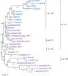
Results: The mean age of the patients was 33 years (SD ± 17.4; 95% CI 22.5 to 43.5 years). Six (46.2%) cases had AK associated risk factors; four patients had ocular trauma and two were contact lens wearers. A. culbertsoni (6/13, 46.2%) was the most common species, followed by A. polyphaga and A. triangularis. Most of the isolates (12/13) belonged to genotype T4 and one was a T12; three sub-clusters T4A, T4B, and T4F were identified within the T4 genotype. There was no significant association between Acanthamoeba types and clinical outcomes. Eight (61.5%) isolates harboured intracellular bacteria and one contained Malassezia restricta. The presence of intracellular microbes was associated with a higher proportion of stromal infiltrates (88.9%, 8/9), epithelial defect (55.6%, 5/9) and hypopyon (55.6%, 5/9) compared to 50% (2/4), 25% (1/4) and 25% (1/4) AK cases without intracellular microbes, respectively.
Conclusions: Genotype T4 was the predominant isolate in southern India. This is the second report of T12 genotype identified from AK patient in India, which is rarely reported worldwide. The majority of the Acanthamoeba clinical isolates in this study harboured intracellular microbes, which may impact clinical characteristics of AK.
Keywords: Acanthamoeba; Endosymbionts; Genotyping; Keratitis.
© 2022. The Author(s).
Conflict of interest statement
The authors declare no competing interests.
Figures


Similar articles
-
The role of naturally acquired intracellular Pseudomonas aeruginosa in the development of Acanthamoeba keratitis in an animal model.PLoS Negl Trop Dis. 2024 Jan 2;18(1):e0011878. doi: 10.1371/journal.pntd.0011878. eCollection 2024 Jan. PLoS Negl Trop Dis. 2024. PMID: 38166139 Free PMC article.
-
Clinical presentations, genotypic diversity and phylogenetic analysis of Acanthamoeba species causing keratitis.J Med Microbiol. 2020 Jan;69(1):87-95. doi: 10.1099/jmm.0.001121. J Med Microbiol. 2020. PMID: 31846414
-
Detection of bacterial endosymbionts in clinical acanthamoeba isolates.Ophthalmology. 2010 Mar;117(3):445-52, 452.e1-3. doi: 10.1016/j.ophtha.2009.08.033. Epub 2010 Jan 19. Ophthalmology. 2010. PMID: 20031220 Free PMC article.
-
Twenty years of acanthamoeba diagnostics in Austria.J Eukaryot Microbiol. 2015 Jan-Feb;62(1):3-11. doi: 10.1111/jeu.12149. Epub 2014 Sep 25. J Eukaryot Microbiol. 2015. PMID: 25047131 Free PMC article. Review.
-
Genotype distribution of Acanthamoeba in keratitis: a systematic review.Parasitol Res. 2021 Sep;120(9):3051-3063. doi: 10.1007/s00436-021-07261-1. Epub 2021 Aug 5. Parasitol Res. 2021. PMID: 34351492 Free PMC article. Review.
Cited by
-
Comparative genomic analysis of Acanthamoeba from different sources and horizontal transfer events of antimicrobial resistance genes.mSphere. 2024 Oct 29;9(10):e0054824. doi: 10.1128/msphere.00548-24. Epub 2024 Oct 1. mSphere. 2024. PMID: 39352766 Free PMC article.
-
Experimental Induction of Acute Acanthamoeba castellanii Keratitis in Cats.Transl Vis Sci Technol. 2023 Aug 1;12(8):10. doi: 10.1167/tvst.12.8.10. Transl Vis Sci Technol. 2023. PMID: 37566398 Free PMC article.
-
Bacterial pathogens and antimicrobial susceptibility in ocular infections: A study at Boru-Meda General Hospital, Dessie, Ethiopia.BMC Ophthalmol. 2024 Aug 13;24(1):342. doi: 10.1186/s12886-024-03544-0. BMC Ophthalmol. 2024. PMID: 39138386 Free PMC article.
-
The role of naturally acquired intracellular Pseudomonas aeruginosa in the development of Acanthamoeba keratitis in an animal model.PLoS Negl Trop Dis. 2024 Jan 2;18(1):e0011878. doi: 10.1371/journal.pntd.0011878. eCollection 2024 Jan. PLoS Negl Trop Dis. 2024. PMID: 38166139 Free PMC article.
-
Zooming in on the intracellular microbiome composition of bacterivorous Acanthamoeba isolates.ISME Commun. 2024 Jan 23;4(1):ycae016. doi: 10.1093/ismeco/ycae016. eCollection 2024 Jan. ISME Commun. 2024. PMID: 38500701 Free PMC article.
References
MeSH terms
Substances
LinkOut - more resources
Full Text Sources
Medical

