Binding of Natural Inhibitors to Respiratory Complex I
- PMID: 36145309
- PMCID: PMC9503403
- DOI: 10.3390/ph15091088
Binding of Natural Inhibitors to Respiratory Complex I
Abstract
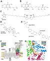
NADH:ubiquinone oxidoreductase (respiratory complex I) is a redox-driven proton pump with a central role in mitochondrial oxidative phosphorylation. The ubiquinone reduction site of complex I is located in the matrix arm of this large protein complex and connected to the membrane via a tunnel. A variety of chemically diverse compounds are known to inhibit ubiquinone reduction by complex I. Rotenone, piericidin A, and annonaceous acetogenins are representatives of complex I inhibitors from biological sources. The structure of complex I is determined at high resolution, and inhibitor binding sites are described in detail. In this review, we summarize the state of knowledge of how natural inhibitors bind in the Q reduction site and the Q access pathway and how their inhibitory mechanisms compare with that of a synthetic anti-cancer agent.
Keywords: NADH dehydrogenase; Parkinson’s disease; acetogenin; mitochondria; piericidin; respiratory chain; rotenone.
Conflict of interest statement
The authors declare no conflict of interest.
Figures


Similar articles
-
Cryo-electron microscopy reveals how acetogenins inhibit mitochondrial respiratory complex I.J Biol Chem. 2022 Mar;298(3):101602. doi: 10.1016/j.jbc.2022.101602. Epub 2022 Jan 19. J Biol Chem. 2022. PMID: 35063503 Free PMC article.
-
Natural substances (acetogenins) from the family Annonaceae are powerful inhibitors of mitochondrial NADH dehydrogenase (Complex I).Biochem J. 1994 Jul 1;301 ( Pt 1)(Pt 1):161-7. doi: 10.1042/bj3010161. Biochem J. 1994. PMID: 8037664 Free PMC article.
-
Ubiquinone Binding and Reduction by Complex I-Open Questions and Mechanistic Implications.Front Chem. 2021 Apr 30;9:672851. doi: 10.3389/fchem.2021.672851. eCollection 2021. Front Chem. 2021. PMID: 33996767 Free PMC article. Review.
-
The reaction sites of rotenone and ubiquinone with mitochondrial NADH dehydrogenase.Biochim Biophys Acta. 1994 Aug 30;1187(2):198-202. doi: 10.1016/0005-2728(94)90110-4. Biochim Biophys Acta. 1994. PMID: 8075112
-
Complex I inhibitors as insecticides and acaricides.Biochim Biophys Acta. 1998 May 6;1364(2):287-96. doi: 10.1016/s0005-2728(98)00034-6. Biochim Biophys Acta. 1998. PMID: 9593947 Review.
Cited by
-
Monoterpenoid Epoxidiol Ameliorates the Pathological Phenotypes of the Rotenone-Induced Parkinson's Disease Model by Alleviating Mitochondrial Dysfunction.Int J Mol Sci. 2023 Mar 19;24(6):5842. doi: 10.3390/ijms24065842. Int J Mol Sci. 2023. PMID: 36982914 Free PMC article.
-
Rotenone Blocks the Glucocerebrosidase Enzyme and Induces the Accumulation of Lysosomes and Autophagolysosomes Independently of LRRK2 Kinase in HEK-293 Cells.Int J Mol Sci. 2023 Jun 24;24(13):10589. doi: 10.3390/ijms241310589. Int J Mol Sci. 2023. PMID: 37445771 Free PMC article.
-
β-lapachone-mediated WST1 Reduction as Indicator for the Cytosolic Redox Metabolism of Cultured Primary Astrocytes.Neurochem Res. 2023 Jul;48(7):2148-2160. doi: 10.1007/s11064-023-03878-z. Epub 2023 Feb 22. Neurochem Res. 2023. PMID: 36811754 Free PMC article.
-
High Yield of Functional Dopamine-like Neurons Obtained in NeuroForsk 2.0 Medium to Study Acute and Chronic Rotenone Effects on Oxidative Stress, Autophagy, and Apoptosis.Int J Mol Sci. 2023 Oct 30;24(21):15744. doi: 10.3390/ijms242115744. Int J Mol Sci. 2023. PMID: 37958728 Free PMC article.
-
Rotenone Induces a Neuropathological Phenotype in Cholinergic-like Neurons Resembling Parkinson's Disease Dementia (PDD).Neurotox Res. 2024 Jun 6;42(3):28. doi: 10.1007/s12640-024-00705-3. Neurotox Res. 2024. PMID: 38842585 Free PMC article.
References
Publication types
Grants and funding
LinkOut - more resources
Full Text Sources
Other Literature Sources

