. 2022 Sep 15;14(18):3812.
doi: 10.3390/nu14183812.

Grifola frondosa Extract Containing Bioactive Components Blocks Skin Fibroblastic Inflammation and Cytotoxicity Caused by Endocrine Disrupting Chemical, Bisphenol A

Affiliations

Grifola frondosa Extract Containing Bioactive Components Blocks Skin Fibroblastic Inflammation and Cytotoxicity Caused by Endocrine Disrupting Chemical, Bisphenol A

Ju-Ha Kim et al. Nutrients. .

Abstract

Grifola frondosa (GF), a species of Basidiomycotina, is widely distributed across Asia and has been used as an immunomodulatory, anti-bacterial, and anti-cancer agent. In the present study, the pharmacological activity of the GF extract against an ecotoxicological industrial chemical, bisphenol A (BPA) in normal human dermal fibroblasts (NHDFs), was investigated. GF extract containing naringin, hesperidin, chlorogenic acid, and kaempferol showed an inhibitory effect on cell death and inflammation induced by BPA in the NHDFs. For the cell death caused by BPA, GF extract inhibited the production of reactive oxygen species responsible for the unique activation of the extracellular signal-regulated kinase. In addition, GF extract attenuated the expression of apoptosis-related proteins (Bax, Bcl-2, and cleaved caspase-3) and the pro-inflammatory cytokine IL-1β by the suppression of the redox-sensitive transcription factor, nuclear factor-kappa B (NF-κB) in BPA-treated NHDFs. For the inflammation triggered by BPA, GF extract blocked the inflammasome-mediated caspase-1 activation that leads to the secretion of IL-1β protein. These results indicate that the GF extract is a functional antioxidant that prevents skin fibroblastic pyroptosis induced by BPA.

Keywords: Grifola frondosa; apoptotic cell death; bisphenol A; normal human dermal fibroblasts; pyroptosis; reactive oxygen species.


Conflict of interest statement

The authors declare no conflict of interest.

Figures

Figure 1
Figure 1
Inhibitory effect of Grifola frondosa (GF) on skin cytotoxicity and inflammation stimulated by bisphenol A (BPA). (A) Dose-dependent response of cell viability in normal human dermal fibroblasts (NHDFs) treated with BPA (0–500 μM) are shown. The cell viability was determined by EZ-CYTOX assay. n = 3. * p ≤ 0.05 vs. 0 μM. (B) Time-dependent response of cell viability treated with BPA (50 μM) is shown. n = 3. * p ≤ 0.05 vs. 0 h. (C) NHDFs were co-treated with GF (100 μg/mL) and BPA for 6 h. n = 4. * p ≤ 0.01 vs. Cont. # p ≤ 0.05 vs. BPA alone. (D) NHDF was treated with BPA for 6 h. The effect of BPA on the expression of pro-inflammatory cytokines was determined by qRT-PCR. * p ≤ 0.01 vs. Cont. n = 3. (E) NHDF was exposed to the BPA in the presence of GF for 6 h. The IL-1β mRNA level is shown. * p ≤ 0.05 versus control. # p ≤ 0.01 vs. BPA alone. n = 3. (F) The level of IL-1β production regulated by GF in BPA-treated NHDF for 6 h was quantified by ELISA. * p ≤ 0.01 vs. Cont. # p ≤ 0.01 vs. BPA alone. n = 3.
Figure 2
Figure 2
GF contains anti-oxidative components that scavenge the intracellular ROS caused by BPA. (A) Time-dependent responses of reactive oxygen species (ROS) production in NHDFs treated with BPA are shown. The level of ROS was determined by staining NHDFs with CM-H2DCFDA. n = 4. * p ≤ 0.05 vs. 0 min. (B) NHDFs were co-treated with GF and BPA for 3 min. n = 4. * p ≤ 0.05 vs. control. # p ≤ 0.01 vs. BPA alone. RFU, Relative fluorescence units. (C) The blocking effects of GF on ROS production (green) confirmed by confocal microscopy are shown. Scale bars, 100 μm (magnification × 100). n = 3. NHDFs were pretreated with 1 μM of N-acetylcysteine (NAC) for 30 min prior to BPA treatment for 6 h. The level of cell proliferation (D) and IL-1β mRNA (E) is shown. * p ≤ 0.01 versus control. # p ≤ 0.01 vs. BPA alone. n = 3.
Figure 3
Figure 3
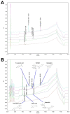
The GF contains flavonol and polyphenolic compounds responsible for its antioxidant properties. (A) UPLC profile of the internal standard compounds. (B) UPLC profile of six major compounds in GF. The insets indicate the chemical structures of naringin, hesperidin, p-coumaric acid, chlorogenic acid, kaempferol, and caffeic acid, respectively.
Figure 4
Figure 4
GF inhibits the phosphorylation of ERK in BPA-induced NHDFs. (A) Time-dependent response of phosphorylation of MAPKs in NHDFs treated with BPA is shown. n = 4. * p ≤ 0.05 vs. 0 min. ROD, relative optical density. (B) The inhibitory effect of GF on phosphorylation of ERK in BPA-treated NHDFs is shown. n = 4. * p ≤ 0.05 vs. Cont., # p ≤ 0.01 vs. BPA alone. (C) NHDFs were incubated with NAC for 30 min prior to BPA treatment for 30 min. n = 4. * p ≤ 0.05 vs. Cont. # p ≤ 0.01 vs. BPA alone. NHDFs were pretreated with ERK inhibitor, PD98059 (1 μM) for 30 min prior to BPA exposure for 6 h. n = 4. The level of cell proliferation (D) and IL-1β mRNA (E) is shown. * p ≤ 0.01 vs. control. # p ≤ 0.01 vs. BPA alone. n = 4. The green squares indicate the cells treated with GF, NAC, or PD98059 alone.
Figure 5
Figure 5
GF inhibits the NF-κB activation to regulate the IL-1β expression triggered by BPA. (A) Time-dependent responses of activation of IκBα and NF-κB in NHDFs exposed by BPA are shown. n = 3. * p ≤ 0.05 vs. 0 min. ROD, relative optical density. (B) The inhibitory effect of GF on activation of NF-κB in BPA-treated NHDFs is shown. n = 4. * p ≤ 0.05 vs. Cont., # p ≤ 0.05 vs. BPA alone. (C) NHDFs were pretreated with PD98059 for 30 min prior to BPA exposure for 60 min. n = 4. * p ≤ 0.05 vs. Cont. # p ≤ 0.01 vs. BPA alone. NHDFs were pretreated with NF-κB inhibitor, Bay 11-7082 (1 μM), for 30 min prior to BPA exposure for 6 h. The level of cell viability (D) and IL-1β mRNA (E) is shown. * p ≤ 0.05 vs. Cont. # p ≤ 0.05 vs. BPA alone. n = 4. The blue squares indicate the cells treated with GF, PD98059, or Bay 11-7082 in the presence of BPA. The green squares indicate the cells treated with GF, PD98059, or Bay 11-7082 alone.
Figure 6
Figure 6
GF blocks dermal fibroblastic apoptosis and inflammation caused by BPA. (A) Time-dependent response of expression of cleaved caspase-3, Bcl-2, and Bax in NHDFs exposed by BPA are shown. n = 3. * p ≤ 0.05 vs. 0 h. ROD, relative optical density. (B) The inhibitory effects of GF on expression of apoptosis-related proteins in BPA-treated NHDFs are shown. n = 4. * p ≤ 0.01 vs. Cont., # p ≤ 0.01 vs. BPA alone. (C) NHDFs were pretreated with Bay 11-7082 for 30 min prior to BPA exposure for 6 h. n = 4. * p ≤ 0.01 vs. Cont. # p ≤ 0.01 vs. BPA alone. (D) NHDFs were incubated with GF for 30 min prior to BPA exposure for 6 h. The apoptotic cell proportion stained by Annexin V/PI was analyzed by performing flow cytometry. n = 4. * p ≤ 0.01 vs. control. (E) The level of ATP production regulated by GF in BPA-treated NHDF for 6 h. * p ≤ 0.05 vs. Cont. # p ≤ 0.01 vs. BPA alone. n = 4. (F) Cells were treated with NAC, PD98059, and Bay11-7082 for 30 min prior to BPA exposure for 6 h. w is shown. * p ≤ 0.01 vs. Cont. # p ≤ 0.05 vs. BPA alone. n = 4. The green squares indicate the cells treated with GF, NAC, PD98059, or Bay 11-7082 alone.
Figure 7
Figure 7
GF inhibits dermal fibroblastic pyroptosis caused by BPA. (A) The inhibitory effect of GF on ASC speck formation (green) confirmed by confocal microscopy is shown. Phalloidin was used for F-actin counterstaining (red). Scale bars, 100 μm (magnification × 400). n = 4. (B) Time-dependent responses of expression of pro- and cleaved caspase-1 exposed by BPA are shown. n = 4. * p ≤ 0.01 vs. 0 h. ROD, relative optical density. (C) The inhibitory effects of GF on expression of pro- and cleaved caspase-1 in BPA-treated NHDFs are shown. n = 4. * p ≤ 0.01 vs. Cont. # p ≤ 0.05 vs. BPA alone. NHDFs were exposed to NAC (D), PD98059 (E), and Bay 11-7082 (F) for 30 min prior to BPA treatment for 6 h. n = 4. * p ≤ 0.05 vs. Cont. # p ≤ 0.01 vs. BPA alone. (G) The sequences of presumed signaling pathways regulated by GF are summarized.

Similar articles

Cited by

References

    1. Cao Y., Qu X., Ming Z., Yao Y., Zhang Y. The correlation between exposure to BPA and the decrease of the ovarian reserve. Int. J. Clin. Exp. Pathol. 2018;11:3375–3382. - PMC - PubMed
    1. Fenichel P., Chevalier N., Brucker-Davis F. Bisphenol A: An endocrine and metabolic disruptor. Ann. Endocrinol. 2013;74:211–220. doi: 10.1016/j.ando.2013.04.002. - DOI - PubMed
    1. Rubin B.S. Bisphenol A: An endocrine disruptor with widespread exposure and multiple effects. J. Steroid Biochem. Mol. Biol. 2011;127:27–34. doi: 10.1016/j.jsbmb.2011.05.002. - DOI - PubMed
    1. Zalko D., Jacques C., Duplan H., Bruel S., Perdu E. Viable skin efficiently absorbs and metabolizes bisphenol A. Chemosphere. 2011;82:424–430. doi: 10.1016/j.chemosphere.2010.09.058. - DOI - PubMed
    1. Geens T., Aerts D., Berthot C., Bourguignon J.P., Goeyens L., Lecomte P., Maghuin-Rogister G., Pironnet A.M., Pussemier L., Scippo M.L., et al. A review of dietary and non-dietary exposure to bisphenol-A. Food Chem. Toxicol. 2012;50:3725–3740. doi: 10.1016/j.fct.2012.07.059. - DOI - PubMed

MeSH terms

Grants and funding

The following are results of a study on the “Leaders in Industry-university Cooperation 3.0” Project, supported by the Ministry of Education and National Research Foundation of Korea and this research also was supported by a National Research Foundation of Korea (NRF) grant funded by the Korean government (MSIT) (2018R1A5A2025272 and 2019R1A2C1088927).