

Sustained Increase in Serum Glial Fibrillary Acidic Protein after First ST-Elevation Myocardial Infarction
- PMID: 36142218
- PMCID: PMC9499398
- DOI: 10.3390/ijms231810304
Sustained Increase in Serum Glial Fibrillary Acidic Protein after First ST-Elevation Myocardial Infarction
Abstract
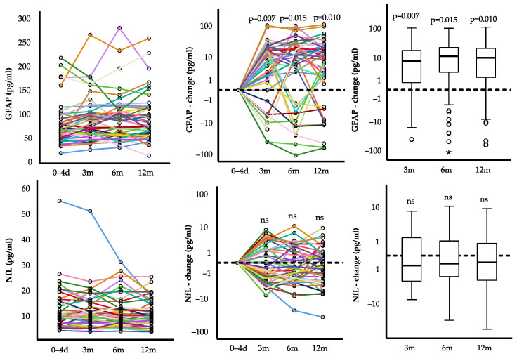
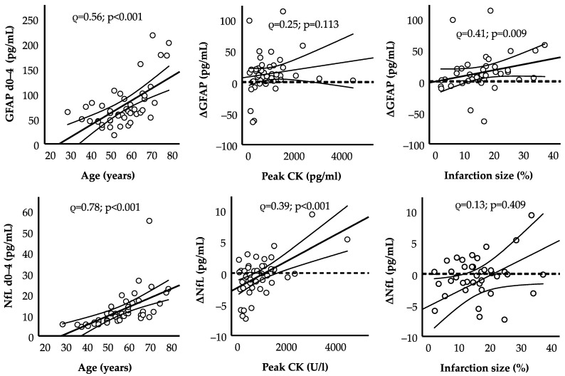
Acute ischemic cardiac injury predisposes one to cognitive impairment, dementia, and depression. Pathophysiologically, recent positron emission tomography data suggest astroglial activation after experimental myocardial infarction (MI). We analyzed peripheral surrogate markers of glial (and neuronal) damage serially within 12 months after the first ST-elevation MI (STEMI). Serum levels of glial fibrillary acidic protein (GFAP) and neurofilament light chain (NfL) were quantified using ultra-sensitive molecular immunoassays. Sufficient biomaterial was available from 45 STEMI patients (aged 28 to 78 years, median 56 years, 11% female). The median (quartiles) of GFAP was 63.8 (47.0, 89.9) pg/mL and of NfL 10.6 (7.2, 14.8) pg/mL at study entry 0-4 days after STEMI. GFAP after STEMI increased in the first 3 months, with a median change of +7.8 (0.4, 19.4) pg/mL (p = 0.007). It remained elevated without further relevant increases after 6 months (+11.7 (0.6, 23.5) pg/mL; p = 0.015), and 12 months (+10.3 (1.5, 22.7) pg/mL; p = 0.010) compared to the baseline. Larger relative infarction size was associated with a higher increase in GFAP (ρ = 0.41; p = 0.009). In contrast, NfL remained unaltered in the course of one year. Our findings support the idea of central nervous system involvement after MI, with GFAP as a potential peripheral biomarker of chronic glial damage as one pathophysiologic pathway.
Keywords: GFAP; MRI; NfL; STEMI; cardiac magnetic resonance imaging; glial damage; glial fibrillary acidic protein; infarction size; myocardial infarction; neurofilament light chain.
Conflict of interest statement
The authors declare no conflict of interest.
Figures
Similar articles
-
Serum glial fibrillary acidic protein and neurofilament light chain in treatment-naïve patients with unipolar depression.J Affect Disord. 2023 Oct 1;338:341-348. doi: 10.1016/j.jad.2023.06.028. Epub 2023 Jun 17. J Affect Disord. 2023. PMID: 37336248
-
Association of serum neurofilament light chain and glial fibrillary acidic protein levels with cognitive decline in Parkinson's disease.Brain Res. 2023 Apr 15;1805:148271. doi: 10.1016/j.brainres.2023.148271. Epub 2023 Feb 7. Brain Res. 2023. PMID: 36754139
-
Neurofilament Light Chain and Glial Fibrillary Acidic Protein as early prognostic biomarkers after out-of-hospital cardiac arrest.Resuscitation. 2023 Dec;193:109983. doi: 10.1016/j.resuscitation.2023.109983. Epub 2023 Sep 29. Resuscitation. 2023. PMID: 37778613
-
Neurofilament light chain as a biomarker in neuromyelitis optica spectrum disorder: a comprehensive review and integrated analysis with glial fibrillary acidic protein.Neurol Sci. 2024 Mar;45(3):1255-1261. doi: 10.1007/s10072-023-07277-8. Epub 2023 Dec 23. Neurol Sci. 2024. PMID: 38141119 Review.
-
Blood Biomarkers as Prognostic Indicators for Neurological Injury in COVID-19 Patients: A Systematic Review and Meta-Analysis.Int J Mol Sci. 2023 Oct 30;24(21):15738. doi: 10.3390/ijms242115738. Int J Mol Sci. 2023. PMID: 37958721 Free PMC article. Review.
Cited by
-
Chronic Neuroinflammation and Cognitive Decline in Patients with Cardiac Disease: Evidence, Relevance, and Therapeutic Implications.Life (Basel). 2023 Jan 24;13(2):329. doi: 10.3390/life13020329. Life (Basel). 2023. PMID: 36836686 Free PMC article. Review.
-
A Molecular (Not Very Becoming) Picture of Stressed Arteries and Heart, with Some Therapeutic Hope.Int J Mol Sci. 2023 Feb 15;24(4):3870. doi: 10.3390/ijms24043870. Int J Mol Sci. 2023. PMID: 36835280 Free PMC article.
References
-
- Disease G.B.D., Injury I., Prevalence C. Global, regional, and national incidence, prevalence, and years lived with disability for 310 diseases and injuries, 1990–2015: A systematic analysis for the Global Burden of Disease Study. Lancet. 2016;388:1545–1602. doi: 10.1016/S0140-6736(16)31678-6. - DOI - PMC - PubMed
-
- Honig A., Lousberg R., Wojciechowski F.L., Cheriex E.C., Wellens H.J., van Praag H.M. Depression following a first heart infarct; similarities with and differences from ‘ordinary’ depression. Ned. Tijdschr. Geneeskd. 1997;141:196–199. - PubMed
MeSH terms
Substances
Grants and funding
LinkOut - more resources
Full Text Sources
Medical
Miscellaneous
