. 2022 Aug 29;10(9):2120.
doi: 10.3390/biomedicines10092120.

Intrathecal Injection of the Secretome from ALS Motor Neurons Regulated for miR-124 Expression Prevents Disease Outcomes in SOD1-G93A Mice

Affiliations

Intrathecal Injection of the Secretome from ALS Motor Neurons Regulated for miR-124 Expression Prevents Disease Outcomes in SOD1-G93A Mice

Marta Barbosa et al. Biomedicines. .

Abstract

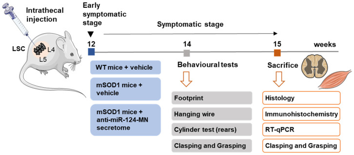
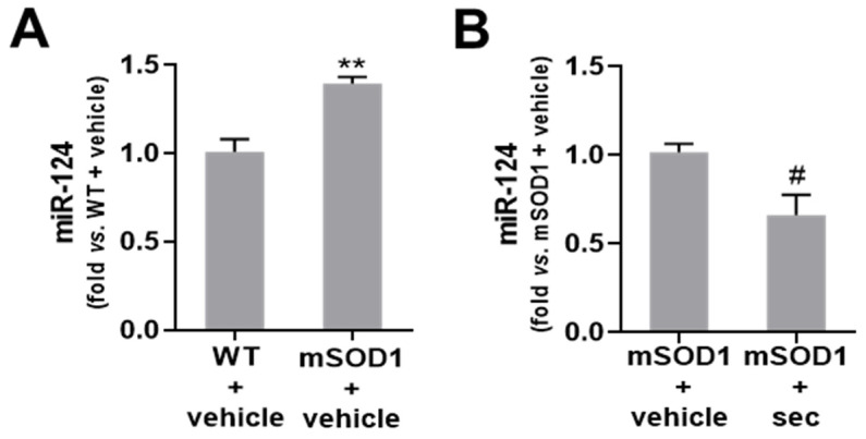
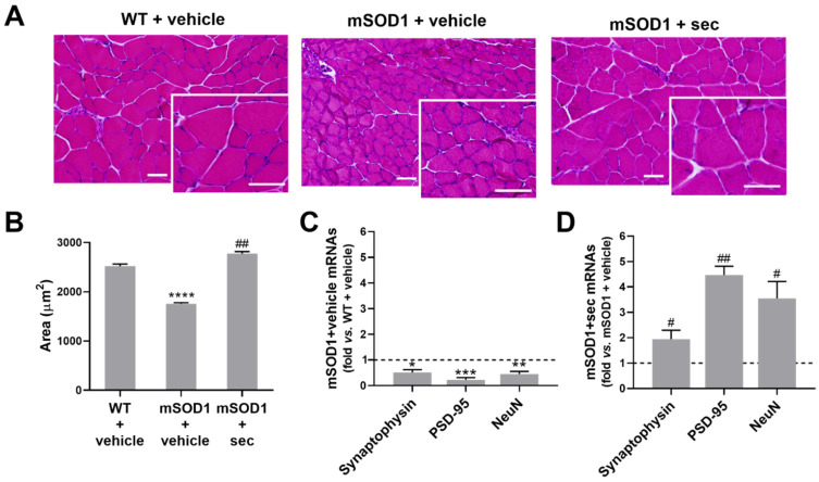
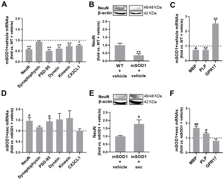
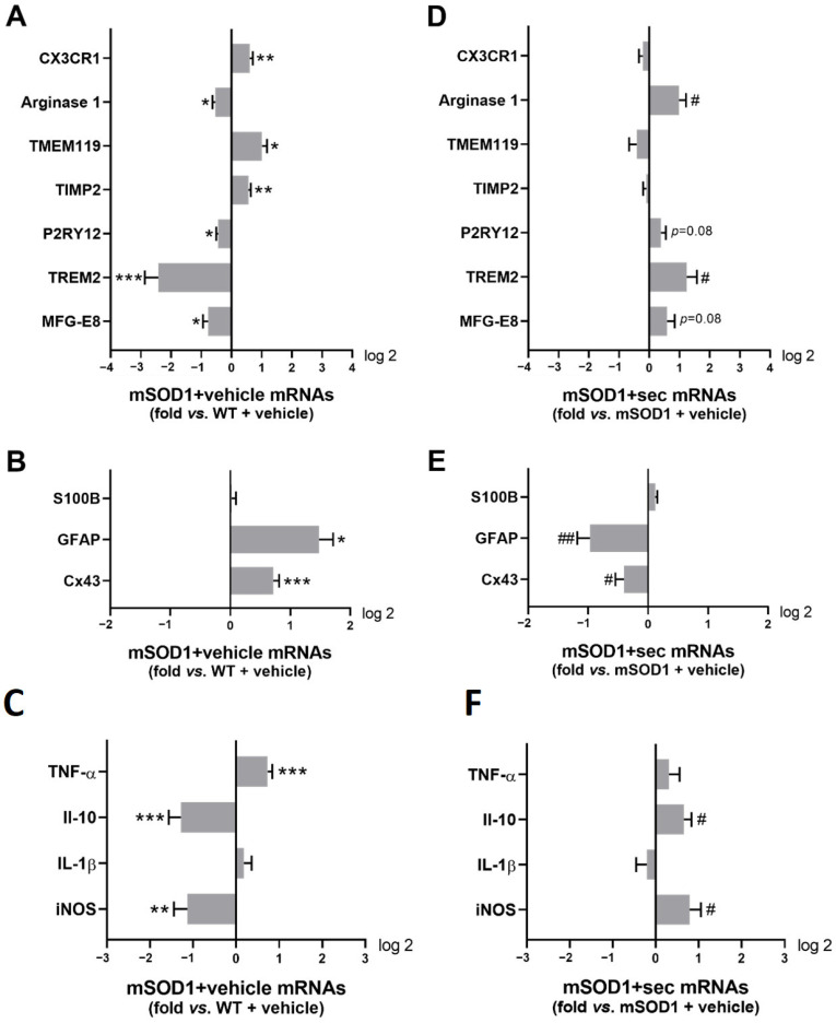
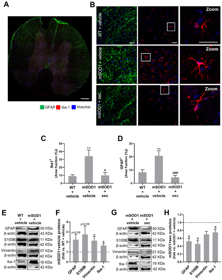
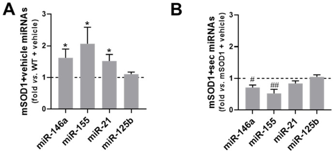
Amyotrophic lateral sclerosis (ALS) is a neurodegenerative disease with short life expectancy and no effective therapy. We previously identified upregulated miR-124 in NSC-34-motor neurons (MNs) expressing human SOD1-G93A (mSOD1) and established its implication in mSOD1 MN degeneration and glial cell activation. When anti-miR-124-treated mSOD1 MN (preconditioned) secretome was incubated in spinal cord organotypic cultures from symptomatic mSOD1 mice, the dysregulated homeostatic balance was circumvented. To decipher the therapeutic potential of such preconditioned secretome, we intrathecally injected it in mSOD1 mice at the early stage of the disease (12-week-old). Preconditioned secretome prevented motor impairment and was effective in counteracting muscle atrophy, glial reactivity/dysfunction, and the neurodegeneration of the symptomatic mSOD1 mice. Deficits in corticospinal function and gait abnormalities were precluded, and the loss of gastrocnemius muscle fiber area was avoided. At the molecular level, the preconditioned secretome enhanced NeuN mRNA/protein expression levels and the PSD-95/TREM2/IL-10/arginase 1/MBP/PLP genes, thus avoiding the neuronal/glial cell dysregulation that characterizes ALS mice. It also prevented upregulated GFAP/Cx43/S100B/vimentin and inflammatory-associated miRNAs, specifically miR-146a/miR-155/miR-21, which are displayed by symptomatic animals. Collectively, our study highlights the intrathecal administration of the secretome from anti-miR-124-treated mSOD1 MNs as a therapeutic strategy for halting/delaying disease progression in an ALS mouse model.

Keywords: ALS mouse model; SOD1-G93A mutation; anti-microRNA-124; intraspinal delivery route; neuroprotection; preservation of motor performance; prevention of glial dysfunction; secretome-based therapy.


Conflict of interest statement

The authors declare no conflict of interest. The funders had no role in the design of the study; in the collection, analyses, or interpretation of data; in the writing of the manuscript; or in the decision to publish the results.

Figures

Figure 1
Figure 1
Experimental design. The wild-type (WT) and SOD1-G93A (mSOD1) mice were injected in the lumbar spinal cord [31] at the early symptomatic stage (12-week-old), either with the MN medium (vehicle, control group) or with the secretome from anti-miR-124-treated mSOD1 MNs (only the mSOD1 mice). Two weeks later, animals were behaviourally characterized through footprint, hanging wire, cylinder, clasping, and grasping tests. At 15 weeks of age, the animals were sacrificed, and the lumbar spinal cord [31] and gastrocnemius muscle were isolated for histological and immunohistological analysis, as well as for reverse transcription quantitative real-time polymerase chain reaction (RT-qPCR) and western blot evaluations. This Figure was partially created with Servier Medical Art (smart.servier.com).
Figure 2
Figure 2
Intrathecal injection of labelled small extracellular vesicles (sEVs) in 12-week-old wild-type (WT) animals leads to their dissemination and interaction with nerve cells, as observed in the lumbar spinal cord (SC) sections at 8 h and 72 h after administration. sEVs were isolated by differential ultracentrifugation and labelled with PKH67 cell linker before injection in the WT mice, which were sacrificed at 8 h and 72 h thereafter. (A,B) Representative images of transversal SC slices with labelled sEVs (green) and respective insets at (A) 8 h and (B) 72 h post-injection. (C,D) Representative images of PKH67-labelled sEVs distributed among (C) NeuN-stained cells and (D) GFAP-stained cells from the lumbar SC of WT mice 72 h after injection. The nuclei were stained with DAPI (blue). DAPI, 4′,6-diamidino-2-phenylindole; GFAP, glial fibrillary acidic protein; NeuN, hexaribonucleotide binding protein 3.
Figure 3
Figure 3
The secretome from mSOD1 MNs transfected with anti-miR-124 abolishes the upregulation of miR-124 in the spinal cord (SC) of ALS mice after 3 weeks of intrathecal injection. Expression of miRNA-124 in the lumbar SC of (A) SOD1-G93A (mSOD1) mice injected at 12 weeks of age with the vehicle (basal media of NSC-34 motor neurons (MNs)) in comparison with the respective wild-type (WT) animals, and (B) mSOD1 mice injected with the secretome derived from anti-miR-124-treated mSOD1 MNs (mSOD1 + sec) in comparison with those injected only with the vehicle. The results were obtained at 15 weeks of age and were normalized to SNORD110. Data are expressed as fold change vs. (A) WT + vehicle and (B) mSOD1 + vehicle (mean ± SEM) from at least 5 animals per group. ** p < 0.01 vs. WT + vehicle; # p < 0.05 vs. mSOD1 + vehicle, unpaired and parametric t-test (with Welch’s correction when needed).
Figure 4
Figure 4
Motor performance, muscular strength, spontaneous activity, and corticospinal function in ALS mice are improved after 2 weeks of intrathecal injection of the secretome from anti-miR-124-treated mSOD1 motor neurons (MNs). Representative illustrations of (A) footprint test, (B) hanging wire test, (C) cylinder test, and (D) clasping/grasping reflexes test. Measurement of the (E) stride length (in centimeters, cm), (F) time holding onto the cage grid (in seconds, s), (G) number of times the mice reared up against the cylinder, and percentage (%) of animals showing the (H) clasping and (I) grasping reflexes in wild-type (WT)/SOD1-G93A (mSOD1) mice injected with the vehicle (basal media of NSC-34 MNs) and mSOD1 mice injected with the secretome derived from mSOD1 MNs modulated with anti-miR-124 (mSOD1 + sec). Data are expressed as mean ± SEM for (EG) and percentage (%) for (H,I) from at least 5 animals per group. **** p < 0.0001, ** p < 0.01 and * p < 0.05 vs. WT + vehicle; #### p < 0.0001 and ## p < 0.01 vs. mSOD1 + vehicle. One-way ANOVA followed by multiple-comparisons Bonferroni post hoc correction was used for footprint and cylinder tests; unpaired and one-way non-parametric ANOVA (Kruskal–Wallis test) was used for hanging wire test; chi-square (and Fisher’s exact) test was used for clasping and grasping tests. Panels (AD) were partially created with Biorender (Biorender.com).
Figure 5
Figure 5
Intrathecal injection of anti-miR-124-treated ALS MN-derived secretome in 12-week-old mSOD1 mice prevents loss of muscle fiber area and the deregulation of genes that direct synaptic proteins at 3 weeks after treatment. (A) Representative images of transversal sections of gastrocnemius muscle from the wild-type (WT) and SOD1-G93A (mSOD1) mice injected with the vehicle (basal media of NSC-34 motor neurons (MNs)), as well as mSOD1 mice injected with the secretome derived from anti-miR-124-treated mSOD1 MNs (mSOD1 + sec), stained with hematoxylin–eosin. Scale bars: 50 µm. (B) Respective average muscle fiber area (in µm). (C) Gene expression of synaptophysin, PSD-95, and NeuN from mSOD1 mice injected with the vehicle in comparison with the respective WT mice, and (D) from mSOD1 mice injected with the secretome in comparison with those injected with the vehicle. Results are mean (±SEM) for (B) and expressed as fold change vs. WT + vehicle for (C) or fold change vs. mSOD1 + vehicle for (D). The images from three fields of the muscle per animal (from three animals per group) were used for histological analysis and five animals per group for RT-qPCR analysis. **** p < 0.0001, *** p < 0.001, ** p < 0.01, and * p < 0.05 vs. WT + vehicle; ## p < 0.01 and # p < 0.05 vs. mSOD1 + vehicle. One-way ANOVA followed by multiple-comparisons Bonferroni post hoc correction was used for (B) and unpaired and parametric t-test with Welch’s correction for (C,D). NeuN, hexaribonucleotide binding protein 3; PSD-95, postsynaptic density protein 95.
Figure 6
Figure 6
Intrathecal injection of the secretome from anti-miR-124-modulated mSOD1 motor neurons (MNs) in the lumbar spinal cord of mSOD1 mice at 12 weeks of age prevents age-associated neurodegeneration after 3 weeks of its administration. (A) Representative images of the ventral horn of the lumbar section grey matter stained by Fluoro-Jade B fluorescence (square) from 15-week-old wild-type (WT)/SOD1-G93A (mSOD1) mice injected with the vehicle (basal media of NSC-34 MNs) and mSOD1 mice injected with the secretome derived from mSOD1 MNs modulated with anti-miR-124 (mSOD1 + sec); (B) the respective quantification of mean fluorescence. Scale bar: 100 µm. Data from 3 animals per group are expressed as fold change (mean ± SEM) vs. WT + vehicle. ** p < 0.01 vs. WT + vehicle; # p < 0.05 vs. mSOD1 + vehicle, one-way ANOVA followed by multiple-comparisons Bonferroni post hoc correction. MFI, mean fluorescence intensity.
Figure 7
Figure 7
Neuronal demise and deficits in synaptic signalling, axonal transport, CX3CL1-CX3CR1 axis, and myelination in the lumbar spinal cord (SC) of 15-week-old mSOD1 mice are prevented by the intrathecal injection of the secretome from modulated mSOD1 motor neurons (MNs) in 12-week-old animals. (A) Gene expression of neuronal-related NeuN, synaptophysin, PSD-95, dynein, kinesin, and CX3CL1; (B) protein expression of NeuN; and (C) myelin-associated genes (MBP, PLP, and GPR17) in the SC of 15-week-old SOD1-G93A (mSOD1) mice that were treated with the vehicle (basal media of NSC-34 MNs) at 12-week-old, in comparison with the respective wild-type (WT) values. (DF) Data from matched experiments realized in mSOD1 mice injected with the secretome derived from anti-miR-124-treated mSOD1 MNs (mSOD1 + sec) in comparison to those treated with the vehicle. (B,E) Representative results from one blot. The results were normalized to RPL-19 for RT-qPCR and β-actin for western blot. Data are expressed as fold change vs. (AC) WT + vehicle and (DF) mSOD1 + vehicle (mean ± SEM) from at least 5 animals per group. ** p < 0.01 and * p < 0.05 vs. WT + vehicle; ## p < 0.01 and # p < 0.05 vs. mSOD1 + vehicle, unpaired and parametric t-test (with Welch’s correction when needed). CX3CL1, C-X3-C motif chemokine ligand 1/fractalkine; GPR17, G-protein-coupled receptor 17; MBP, myelin basic protein; NeuN, hexaribonucleotide-binding protein 3; PLP, myelin proteolipid protein; PSD-95, postsynaptic density protein 95; RPL19, 60S ribosomal L19; RT-qPCR, reverse transcription quantitative real-time polymerase chain reaction.
Figure 8
Figure 8
Intrathecal injection of the secretome from anti-miR-124-modulated mSOD1 motor neurons (MNs) in the lumbar spinal cord (SC) of mSOD1 mice at 12-week-old prevents microglia activation, astrocyte reactivity, TNF-α signalling, and inflammation associated with the symptomatic stage. Gene expression of (A) microglia-associated markers (MFG-E8, TREM 2, P2RY12, TIMP2, arginase 1, and CX3CR1); (B) astrocyte-related markers (Cx43, GFAP, and S100B); and (C) inflammatory-associated markers (iNOS, IL-1β, IL-10, and TNF-α) in the SC of SOD1-G93A (mSOD1) mice injected with the vehicle (basal media of NSC-34 MNs) in comparison to those in wild-type (WT) animals; (DF) after the administration of the secretome derived from anti-miR-124-treated mSOD1 MNs (mSOD1 + sec) as compared with mSOD1 mice treated with the vehicle. The results were normalized to RPL-19. Data are expressed as fold change vs. (AC) WT + vehicle and (DF) mSOD1 + vehicle (mean ± SEM) from at least 5 animals per group. *** p < 0.001, ** p < 0.01, and * p < 0.05 vs. WT + vehicle; ## p < 0.01 and # p < 0.05 vs. mSOD1 + vehicle, unpaired and parametric t-test (with Welch’s correction when needed). CX3CR1, c-x3-c chemokine receptor 1; Cx43, connexin 43; GFAP, glial fibrillary acidic protein; IL-10, interleukin 10; IL-1β, interleukin 1β; iNOS, inducible nitric oxide synthase; MFG-E8, milk fat globule epidermal growth factor 8; P2RY12, purinergic receptor p2y12; RPL19, 60S ribosomal L19; S100B, S100 calcium-binding protein B; TIMP2, tissue inhibitor of metalloproteinases 2; TMEM119, transmembrane protein 119; TNF-α, tumour necrosis factor alpha; TREM2, triggering receptor expressed on myeloid cells 2.
Figure 9
Figure 9
Intrathecal injection of the secretome from anti-miR-124-modulated mSOD1 motor neurons (MNs) in the lumbar spinal cord (SC) of mSOD1 mice at 12-week-old counteracts the upregulation of proteins linked to glia-driven immunoreactivity processes after 3 weeks of its administration. (A) Representative image of a transversal SC slice stained with Iba-1 (microglia-associated marker) and glial fibrillary acidic protein/GFAP (astrocyte-associated marker). (B) Representative images for GFAP- (green) and Iba-1- (red) positive cells from the lumbar SC of WT and SOD1-G93A (mSOD1) mice injected with the vehicle (basal media of NSC-34 MNs) and mSOD1 mice injected with the secretome derived from anti-miR-124-treated mSOD1 MNs (mSOD1 + sec). A zoomed-in image of an Iba-1-positive cell is shown. Nuclei were stained with Hoechst (blue). Scale bar: (A) 300 µm and (B) 30 µm. (C,D) Area fraction (in percentage, %) occupied by GFAP- and Iba-1-positive cells, respectively. (E,G) Representative western blots (WB). (F,H) Data resulting from WB analysis of reactive astrocytic markers (GFAP, S100B, and vimentin) and microglial Iba-1. β-actin was used as a loading control for WB analysis. Results are mean (± SEM) for (C,D) and expressed as fold change vs. WT + vehicle for (F) or fold change vs. mSOD1 + vehicle for (H). The images were analysed from five ventral horn fields per animal (from three animals per group) for immunohistochemistry and five animals per group for WB analysis. ** p < 0.01 and * p < 0.05 vs. WT + vehicle; ### p < 0.001, and # p < 0.05 vs. mSOD1 + vehicle. One-way ANOVA followed by multiple-comparisons Bonferroni post hoc correction was used for (C,D), and unpaired and parametric t-test with Welch’s correction for (F,H). GFAP, glial fibrillary acidic protein; Iba-1, ionized calcium-binding adaptor molecule 1; S100B, S100 calcium-binding protein B.
Figure 10
Figure 10
Upregulation of inflammatory-associated miRNA observed in the lumbar spinal cord (SC) of ALS mice is prevented by the secretome from the mSOD1 MNs engineered with anti-miR-124. Expression of inflammatory-associated micro(mi)RNAs (miR-146, miR-155, miR-21, miR-124, and miR-125b) in the SC of (A) SOD1-G93A (mSOD1) mice injected with the vehicle (basal media of NSC-34 motor neurons (MNs)) in comparison with those in wild-type (WT) animals, and (B) mSOD1 mice injected with the secretome derived from anti-miR-124-treated mSOD1 MNs (mSOD1 + sec) in comparison with those treated with the vehicle. The results were normalized to SNORD110. Data are expressed as fold change vs. (A) WT + vehicle and (B) mSOD1 + vehicle (mean ± SEM) from at least 5 animals per group. * p < 0.05 vs. WT + vehicle; ## p < 0.01 and # p < 0.05 vs. mSOD1 + vehicle, unpaired and parametric t-test (with Welch’s correction when needed).

Similar articles

Cited by

References

    1. Hardiman O., Al-Chalabi A., Chio A., Corr E.M., Logroscino G., Robberecht W., Shaw P.J., Simmons Z., van den Berg L.H. Amyotrophic lateral sclerosis. Nat. Rev. Dis. Primers. 2017;3:17071. doi: 10.1038/nrdp.2017.71. - DOI - PubMed
    1. Vahsen B.F., Gray E., Thompson A.G., Ansorge O., Anthony D.C., Cowley S.A., Talbot K., Turner M.R. Non-neuronal cells in amyotrophic lateral sclerosis—From pathogenesis to biomarkers. Nat. Rev. Neurol. 2021;17:333–348. doi: 10.1038/s41582-021-00487-8. - DOI - PubMed
    1. Xie M., Zhao S., Bosco D.B., Nguyen A., Wu L.J. Microglial TREM2 in amyotrophic lateral sclerosis. Dev. Neurobiol. 2022;82:125–137. doi: 10.1002/dneu.22864. - DOI - PMC - PubMed
    1. Rosen D.R., Siddique T., Patterson D., Figlewicz D.A., Sapp P., Hentati A., Donaldson D., Goto J., O’Regan J.P., Deng H.X., et al. Mutations in Cu/Zn superoxide dismutase gene are associated with familial amyotrophic lateral sclerosis. Nature. 1993;362:59–62. doi: 10.1038/362059a0. - DOI - PubMed
    1. Alrafiah A.R. From Mouse Models to Human Disease: An Approach for Amyotrophic Lateral Sclerosis. In Vivo. 2018;32:983–998. doi: 10.21873/invivo.11339. - DOI - PMC - PubMed

LinkOut - more resources