

Ultra-Morphology and Mechanical Function of the Trichoideum Sensillum in Nabis rugosus (Linnaeus, 1758) (Insecta: Heteroptera: Cimicomorpha)
- PMID: 36135500
- PMCID: PMC9504417
- DOI: 10.3390/insects13090799
Ultra-Morphology and Mechanical Function of the Trichoideum Sensillum in Nabis rugosus (Linnaeus, 1758) (Insecta: Heteroptera: Cimicomorpha)
Abstract
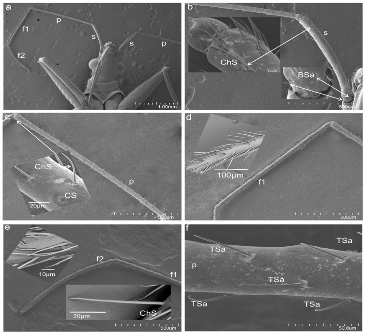
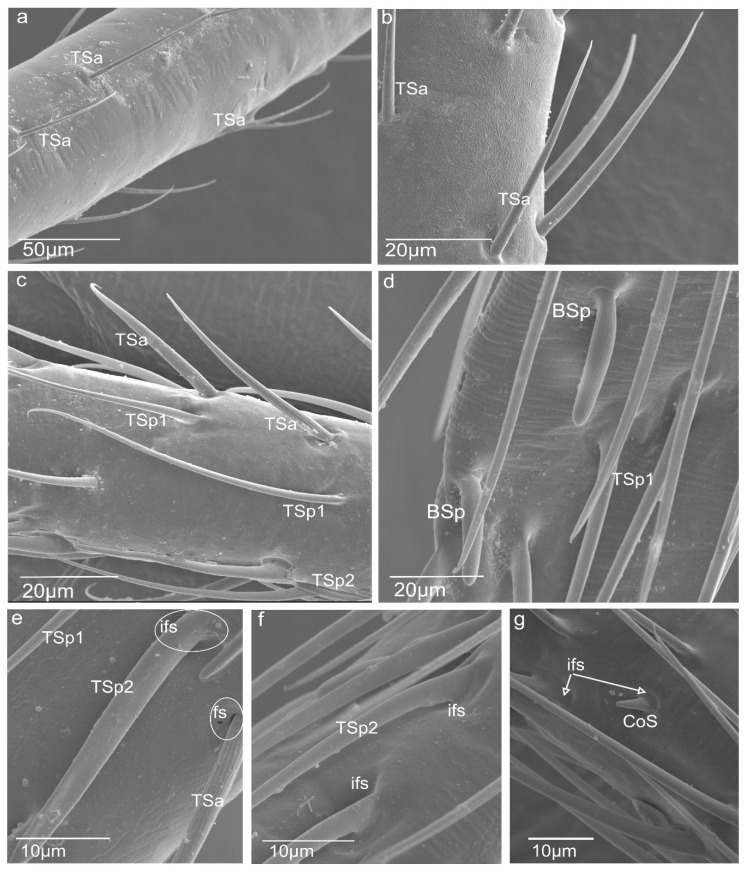
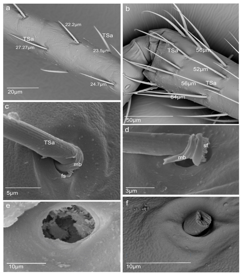
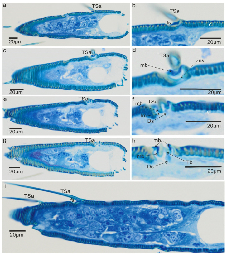
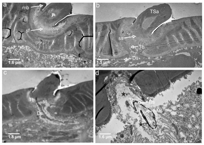
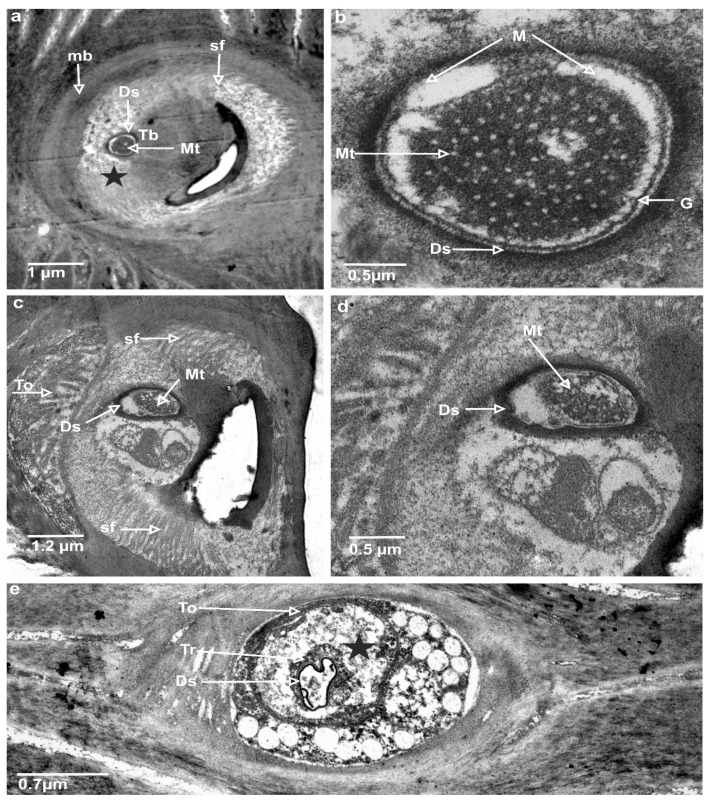
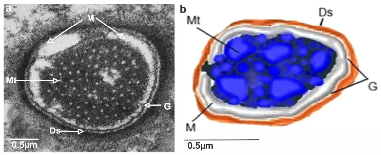
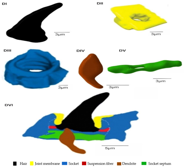
The present study aims to investigate the morphological features of the antennal sensilla by using SEM and TEM. The construction of a 3D model of trichoideum sensillum using Amira software is presented in this paper. Five sensillum types, namely trichoideum, chaeticum, campaniformium, coeloconicum, and basiconicum, were recorded. This model exhibits the mechanosensillum components, including the embedded hair in a socket attached by the joint membrane and the dendrite connected to the hair base passing through the cuticle layers. TEM images present the dendrite way, micro-tubules inside the dendritic sheath, and terminal structure of the tubular dendrite body and so-called companion cells included in the receptor, e.g., tormogen and trichogen. The parameters noted for the external structure and ultrastructure of the mechano-receptor indicate that they are specific to a particular type of sensillum and would be useful in developing the model for a biosensor. Results show that bio-inspired sensors can be developed based on morphological and ultrastructural studies and to conduct mechanical studies on their components.
Keywords: 3D model; N. rugosus; mechanoreceptor; morphology; scanning electron microscopy; transmission electron microscopy; trichoideum sensillum.
Conflict of interest statement
The authors declare no conflict of interest.
Figures
Similar articles
-
Ultrastructure of a Mechanoreceptor of the Trichoid Sensilla of the Insect Nabis rugosus: Stimulus-Transmitting and Bio-Sensory Architecture.Bioengineering (Basel). 2023 Jan 11;10(1):97. doi: 10.3390/bioengineering10010097. Bioengineering (Basel). 2023. PMID: 36671669 Free PMC article.
-
Three-dimensional architecture of a mechanoreceptor in the brown planthopper, Nilaparvata lugens, revealed by FIB-SEM.Cell Tissue Res. 2020 Mar;379(3):487-495. doi: 10.1007/s00441-019-03122-7. Epub 2019 Nov 25. Cell Tissue Res. 2020. PMID: 31768711
-
Ultrastructure of antennal sensilla of the peach aphid Myzus persicae Sulzer, 1776.J Morphol. 2015 Feb;276(2):219-27. doi: 10.1002/jmor.20335. Epub 2014 Nov 4. J Morphol. 2015. PMID: 25366941
-
Functional morphology of insect mechanoreceptors.Microsc Res Tech. 1997 Dec 15;39(6):506-31. doi: 10.1002/(SICI)1097-0029(19971215)39:6<506::AID-JEMT5>3.0.CO;2-B. Microsc Res Tech. 1997. PMID: 9438251 Review.
-
Fine structure of olfactory sensilla in myriapods and arachnids.Microsc Res Tech. 1992 Sep 1;22(4):372-91. doi: 10.1002/jemt.1070220406. Microsc Res Tech. 1992. PMID: 1392066 Review.
Cited by
-
Functional Morphology and Ultrastructure of the Peripheral Antennal Sensillar System of Graphosoma italicum (Müller, 1766) (Insecta: Hemiptera: Pentatomidae).Insects. 2024 Jul 12;15(7):528. doi: 10.3390/insects15070528. Insects. 2024. PMID: 39057261 Free PMC article.
-
Closer view of antennal sensory organs of two Leptoglossus species (Insecta, Hemiptera, Coreidae).Sci Rep. 2023 Jan 12;13(1):617. doi: 10.1038/s41598-023-27837-4. Sci Rep. 2023. PMID: 36635483 Free PMC article.
-
Ultrastructure of a Mechanoreceptor of the Trichoid Sensilla of the Insect Nabis rugosus: Stimulus-Transmitting and Bio-Sensory Architecture.Bioengineering (Basel). 2023 Jan 11;10(1):97. doi: 10.3390/bioengineering10010097. Bioengineering (Basel). 2023. PMID: 36671669 Free PMC article.
References
-
- Kerzhner I.M. In: Poluzhestkokrylye Semeystva Nabidae [Heteroptera of the Family Zoo Morphology] 2nd ed. Theodor O., editor. Fauna of the USSR; Leningrad, Russia: 1981. pp. 1–326.
-
- Kerzhner I.M. Family Nabidae, A. Costa—damsel bugs, 1853. In: Aukema B., Rieger C., editors. Catalogue of the Heteroptera of the Palaearctic Region. 2nd ed. The Netherlands Entomological Society; Wageningen, The Netherlands: 2004. pp. 84–107.
-
- Brożek J. Morphology and arrangement of the labial sensilla of the water bugs. Bull. Insectol. 2008;61:275–297. doi: 10.1007/s00435-012-0174-z. - DOI
-
- Calderón L., Alberto B. Ph.D. Thesis. Eberhard Karls Universität Tübingen; Tübinge, Germany: 2019. The Function of Mechanosensory SYSTEMS in the Startle Behavior of Planktonic Larvae.
Grants and funding
LinkOut - more resources
Full Text Sources
