

Viability and Adhesion of Periodontal Ligament Fibroblasts on a Hydroxyapatite Scaffold Combined with Collagen, Polylactic Acid-Polyglycolic Acid Copolymer and Platelet-Rich Fibrin: A Preclinical Pilot Study
- PMID: 36135161
- PMCID: PMC9497794
- DOI: 10.3390/dj10090167
Viability and Adhesion of Periodontal Ligament Fibroblasts on a Hydroxyapatite Scaffold Combined with Collagen, Polylactic Acid-Polyglycolic Acid Copolymer and Platelet-Rich Fibrin: A Preclinical Pilot Study
Abstract
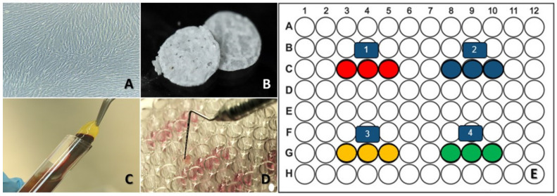
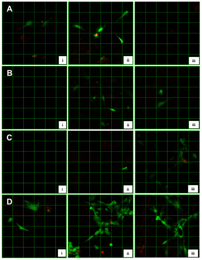
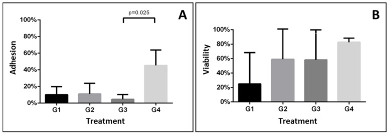
Background: Conventional periodontal therapy relies on bone regeneration strategies utilizing scaffolds made of diverse materials, among which collagen, to promote cell adhesion and growth. Objective: To evaluate periodontal ligament fibroblast (HPdLF) cell adhesion and viability for periodontal regeneration purposes on hydroxyapatite scaffolds containing collagen (HAp-egg shell) combined with polylactic acid−polyglycolic acid copolymer (PLGA) and Platelet-Rich Fibrin (PRF). Methods: Four variations of the HAp-egg shell were used to seed HPdLF for 24 h and evaluate cell viability through a live/dead assay: (1) (HAp-egg shell/PLGA), (2) (HAp-egg shell/PLGA + collagen), (3) (HAp-egg shell/PLGA + PRF) and (4) (HAp-egg shell/PLGA + PRF + collagen). Cell adhesion and viability were determined using confocal microscopy and quantified using central tendency and dispersion measurements; significant differences were determined using ANOVA (p < 0.05). Results: Group 1 presented low cell viability and adhesion (3.70−10.17%); groups 2 and 3 presented high cell viability and low cell adhesion (group 2, 59.2−11.1%, group 3, 58−4.6%); group 4 presented the highest cell viability (82.8%) and moderate cell adhesion (45%) (p = 0.474). Conclusions: The effect of collagen on the HAp-egg shell/PLGA scaffold combined with PRF favored HPdLF cell adhesion and viability and could clinically have a positive effect on bone defect resolution and the regeneration of periodontal ligament tissue.
Keywords: collagen; fibroblasts; hydroxyapatite; periodontal ligament; platelet-rich fibrin; polylactic acid–polyglycolic acid copolymer; tissue engineering.
Conflict of interest statement
The authors declare no conflict of interest.
Figures
Similar articles
-
Accelerating bone regeneration using poly(lactic-co-glycolic acid)/hydroxyapatite scaffolds containing duck feet-derived collagen.Int J Biol Macromol. 2023 Feb 28;229:486-495. doi: 10.1016/j.ijbiomac.2022.12.296. Epub 2022 Dec 29. Int J Biol Macromol. 2023. PMID: 36587641
-
The effect of platelet-rich fibrin exudate addition to porous poly(lactic-co-glycolic acid) scaffold in bone healing: An in vivo study.J Biomed Mater Res B Appl Biomater. 2020 May;108(4):1304-1310. doi: 10.1002/jbm.b.34478. Epub 2019 Aug 19. J Biomed Mater Res B Appl Biomater. 2020. PMID: 31429195
-
Study of platelet-rich fibrin combined with rat periodontal ligament stem cells in periodontal tissue regeneration.J Cell Mol Med. 2018 Feb;22(2):1047-1055. doi: 10.1111/jcmm.13461. J Cell Mol Med. 2018. PMID: 29368432 Free PMC article.
-
Inorganic apatite nanomaterial: Modified surface phenomena and its role in developing collagen based polymeric bio-composite (Coll-PLGA/HAp) for biological applications.Colloids Surf B Biointerfaces. 2018 Dec 1;172:734-742. doi: 10.1016/j.colsurfb.2018.09.038. Epub 2018 Sep 18. Colloids Surf B Biointerfaces. 2018. PMID: 30248644
-
Poly(lactide-co-glycolide)/hydroxyapatite nanofibrous scaffolds fabricated by electrospinning for bone tissue engineering.J Mater Sci Mater Med. 2011 Aug;22(8):1873-84. doi: 10.1007/s10856-011-4374-8. Epub 2011 Jun 18. J Mater Sci Mater Med. 2011. PMID: 21681656
Cited by
-
Navigating the combinations of platelet-rich fibrin with biomaterials used in maxillofacial surgery.Front Bioeng Biotechnol. 2024 Oct 7;12:1465019. doi: 10.3389/fbioe.2024.1465019. eCollection 2024. Front Bioeng Biotechnol. 2024. PMID: 39434715 Free PMC article. Review.
-
Electrospun Scaffolds of Polylactic Acid, Collagen, and Amorphous Calcium Phosphate for Bone Repair.Pharmaceutics. 2023 Oct 25;15(11):2529. doi: 10.3390/pharmaceutics15112529. Pharmaceutics. 2023. PMID: 38004509 Free PMC article.
-
Enhancing orthodontic treatment control with fish scale-derived hydroxyapatite nanoparticles: Insights from an animal model study.Saudi Dent J. 2024 Aug;36(8):1128-1134. doi: 10.1016/j.sdentj.2024.06.007. Epub 2024 Jun 4. Saudi Dent J. 2024. PMID: 39176163 Free PMC article.
References
-
- Rosales-Ibáñez R., Alvarado-Estrada K.N., Ojeda-Gutiérrez F. Tissue Engineering in Dentistry. Rev. ADM. 2012;69:164–167.
-
- Mishra M., Mishra P., Shambharkar V., Raut A. Scaffolds in Periodontal Regeneration. J. Pharm. Biomed. Sc. 2016;6:10–17.
-
- Parra M., Haidar Z.S., Olate S. Use of PRF in Combination with Synthetic Filling Materials (HA and ß-TCP) for Bone Reconstructions. Avances en Odontoestomatologí-a. 2018;34:79–86.
Grants and funding
LinkOut - more resources
Full Text Sources
