. 2023 May;19(5):1406-1423.
doi: 10.1080/15548627.2022.2127240. Epub 2022 Oct 13.

VAMP724 and VAMP726 are involved in autophagosome formation in Arabidopsis thaliana

Affiliations

VAMP724 and VAMP726 are involved in autophagosome formation in Arabidopsis thaliana

Yilin He et al. Autophagy. 2023 May.

Abstract

Macroautophagy/autophagy, an evolutionarily conserved degradative process essential for cell homeostasis and development in eukaryotes, involves autophagosome formation and fusion with a lysosome/vacuole. The soluble N-ethylmaleimide-sensitive factor attachment protein receptor (SNARE) proteins play important roles in regulating autophagy in mammals and yeast, but relatively little is known about SNARE function in plant autophagy. Here we identified and characterized two Arabidopsis SNAREs, AT4G15780/VAMP724 and AT1G04760/VAMP726, involved in plant autophagy. Phenotypic analysis showed that mutants of VAMP724 and VAMP726 are sensitive to nutrient-starved conditions. Live-cell imaging on mutants of VAMP724 and VAMP726 expressing YFP-ATG8e showed the formation of abnormal autophagic structures outside the vacuoles and compromised autophagic flux. Further immunogold transmission electron microscopy and electron tomography (ET) analysis demonstrated a direct connection between the tubular autophagic structures and the endoplasmic reticulum (ER) in vamp724-1 vamp726-1 double mutants. Further transient co-expression, co-immunoprecipitation and double immunogold TEM analysis showed that ATG9 (autophagy related 9) interacts and colocalizes with VAMP724 and VAMP726 in ATG9-positive vesicles during autophagosome formation. Taken together, VAMP724 and VAMP726 regulate autophagosome formation likely working together with ATG9 in Arabidopsis.Abbreviations: ATG, autophagy related; BTH, benzo-(1,2,3)-thiadiazole-7-carbothioic acid S-methyl ester; Conc A, concanamycin A; EM, electron microscopy; ER, endoplasmic reticulum; FRET, Förster/fluorescence resonance energy transfer; MS, Murashige and Skoog; MVB, multivesicular body; PAS, phagophore assembly site; PM, plasma membrane; PVC, prevacuolar compartment; SNARE, soluble N-ethylmaleimide-sensitive factor attachment protein receptor; TEM, transmission electron microscopy; TGN, trans-Golgi network; WT, wild-type.

Keywords: ATG9; Arabidopsis; SNARE; VAMP724; VAMP726; autophagy.


Conflict of interest statement

No potential conflict of interest was reported by the author(s).

Figures
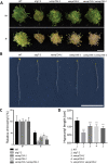
Figure 1.
Figure 1.
The VAMP724 and VAMP726 knock-out (KO) mutants display sensitive phenotypes under nutrient-deficient conditions. (A) vamp724-1 or vamp726-1 mutants showed enhanced sensitivity to nitrogen starvation. 6-day-old seedlings of WT and various indicated mutants from MS plate (+N) were transferred to fresh MS medium (+N) or N-deficient (–N) medium for 2 weeks before photography. Bar: 1 cm. (B) Phenotype of suppressed hypocotyl elongation. Seedlings of WT and various indicated mutants were grown in continuous darkness on MS solid medium without sucrose (-C) for 1 week before photography. Representative photographs of the etiolated seedlings are shown. Bar: 1 cm. (C) Relative chlorophyll contents of seedlings from WT and various mutants shown in (A). Each seedling bundle contained 30 seedlings and the results were obtained from three independent experiments (error bars ± SD). *P < 0.05, **P < 0.01, ***P < 0.001, two-sided Student’s t-test. (D) Quantification of hypocotyl length shown in (B). Results were obtained from three independent experiments of 10 seedlings each (error bars ± SD). *P < 0.05, **P < 0.01, ***P < 0.001, two-sided Student’s t-test.
Figure 2.
Figure 2.
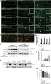
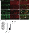
Depletion of VAMP724 and VAMP726 affects the autophagy pathway in Arabidopsis. (A) Detection of YFP-ATG8e-positive autophagic tubules in vamp724-1 and vamp726-1 mutants upon BTH-induced autophagy. 5-day-old seedlings of WT, as well as vamp724-1 and vamp726-1 mutants expressing YFP-ATG8e were incubated in medium without BTH (Left panel, Control), with BTH (Middle panel) or with BTH+Conc A (Right panel) for 6 h, followed by confocal imaging. Arrows indicated examples of the ATG8e-positive tubular structures with enlargement in the insets, while arrowheads indicated examples of autophagic bodies inside the vacuoles. Bar: 10 μm. (B) Quantification of the numbers of YFP-ATG8e-positive autophagosomes in BTH-treated or untreated WT, vamp724-1, vamp726-1, and vamp724-1 vamp726-1 shown in (A). Results were obtained from three independent experiments (error bars ± SD). *P < 0.05, **P < 0.01, two-sided Student’s t-test. (C) Quantification of the numbers of YFP-ATG8e-positive tubular structures in WT, vamp724-1, vamp726-1, and vamp724-1 vamp726-1 upon BTH treatments shown in (A). Results were obtained from three independent experiments (error bars ± SD). *P < 0.05, **P < 0.01, two-sided Student’s t-test. (D) Quantification of the numbers of autophagic bodies inside the vacuoles of WT, vamp724-1, vamp726-1, and vamp724-1 vamp726-1 upon BTH+Conc A treatments shown in (A). Results were obtained from three independent experiments (error bars ± SD). *P < 0.05, ns: not significant (P > 0.05), two-sided Student’s t-test. (E) 5-day-old seedlings of YFP-ATG8e and YFP-ATG8e vamp724-1 vamp726-1 were incubated in MS medium without nitrogen (N-) for 24 h in dark, followed by FM 4-64 uptake for 1 h and subsequent Conc A treatment for 6 h prior to confocal imaging. v, vacuole. Bar: 10 μm. (F) Immunoblot detection of vacuolar turnover of YFP-ATG8e WT and YFP-ATG8e vamp724-1 vamp726-1 upon BTH-induced autophagy. Five-day-old seedlings of YFP-ATG8e and YFP-ATG8e vamp724-1 vamp726-1 were subjected to BTH treatments, followed by total protein extraction at indicated time points and subsequent immunoblot analysis with GFP antibodies whereas cFBPase antibodies were used as a loading control. For quantification analysis, protein band intensities were quantified using ImageJ software and all values were normalized according to the protein loading control (cFBPase) before calculation of vacuolar import ratio (YFP:total). Relative vacuolar import ratio of different time points was normalized to time 0 (which is set to 1). (G) Immunoblot detection of the ATG8 lipidation levels in WT, vamp724-1, vamp726-1, vamp724-1 vamp726-1, and atg5-1. 5-day-old seedlings of WT and various mutants were incubated in medium without (control) or with BTH and Conc A for 6 h, followed by extraction of membrane proteins fractions and subsequent immunoblot analysis with ATG8 antibodies, whereas cFBPase antibodies were used as a loading control.
Figure 3.
Figure 3.
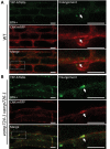
Autophagosome-related tubular structures are associated with the ER membranes in vamp724-1 vamp726-1 double mutant. (A) YFP-ATG8e-positive structures are formed in a close proximity to the ER membrane in WT. 5-day-old YFP-ATG8e CNX-mRFP seedlings were incubated in MS medium with BTH for 6 h before confocal imaging analysis. The dashed square regions are enlarged and shown respectively in the right panels. Arrows indicated examples of association between autophagosome and the ER marker. (B) YFP-ATG8e-positive tubules are accompanied with the ER membrane in vamp724-1 vamp726-1 double mutant. 5-day-old YFP-ATG8e CNX-mRFP vamp724-1 vamp726-1 seedlings were incubated in MS medium with BTH for 6 h before confocal imaging analysis. The dashed square regions are enlarged and shown respectively in the right panels. Arrows indicated examples of association between the YFP-ATG8e-positive tubules and the ER. Bar: 10 μm.
Figure 4.
Figure 4.
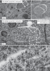
Immunogold-TEM analysis of tubular autophagic structures in root cells of YFP-ATG8e vamp724-1 vamp726-1 upon autophagy induction. Ultrathin sections were prepared from high-pressure frozen/freeze-substituted root cells of YFP-ATG8e vamp724-1 vamp726-1 seedlings upon 6 h BTH treatment, followed by immunogold labeling with indicated antibodies. (A) An overview of BTH-treated root cells of transgenic YFP-ATG8e vamp724-1 vamp726-1 seedlings. Open arrows indicate examples of the tubular autophagic structures. Bar: 2 μm. (B) Enlargement of the dashed box shown in (A). (C) Double immunogold labeling with anti-ATG8 and anti-calreticulin antibodies on autophagosome-related tubular structures in BTH-treated root cells of transgenic YFP-ATG8e vamp724-1 vamp726-1 seedlings. Bar: 500 nm. (D and E) Enlargement of the dashed boxes shown in (C). Arrowheads and arrows indicate gold particles for anti-ATG8 (10 nm) and anti-calreticulin (6 nm), respectively.
Figure 5.
Figure 5.
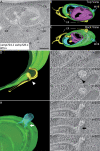
A 3D Electron tomography (ET) analysis reveals direct connection between the autophagic tubule and the ER membranes in root cells of vamp724-1 vamp726-1 mutant upon autophagy induction. A 3D tomographic analysis was performed on the high-pressure frozen root tip cells of vamp724-1 vamp726-1 mutant after 6-h BTH treatment. (A) Tomographic slice showing a representative example of the abnormal autophagic structure in BTH-treated root cells of vamp724-1 vamp726-1 mutant. (B and C) The 3D tomographic models of the autophagic structure shown in (A). (D to G) The 3D model and tomographic slices showing direct connection between the autophagic tubule and the ER as indicated by the arrowhead. (H to K) The 3D model and tomographic slices showing connection between MVB and autophagosome as indicated by the arrow. ER, endoplasmic reticulum; G, Golgi apparatus. MVB, multivesicular body. Bar: 500 nm.
Figure 6.
Figure 6.
VAMP724 and VAMP726 partially colocalized with the autophagosome marker ATG8e upon autophagy induction in transgenic Arabidopsis plants. Transgenic Arabidopsis plants expressing YFP-VAMP724 or YFP-VAMP726 were crossed into the autophagosome marker line mCherry-ATG8e to generate the double transgenic plants of YFP-VAMP724 mCherry-ATG8e and YFP-VAMP726 mCherry-ATG8e respectively. Root cells of 5-day-old double transgenic seedlings were incubated in medium without (A) or with BTH (B) to induce autophagy, followed by confocal imaging analysis. Arrows indicated examples of colocalization between the two fusion proteins with enlargement in the insets. Bar: 10 μm. (C) Quantification of the colocalization ratio of VAMP724 or VAMP726-colocalized autophagosomes over total autophagosomes shown in (A) and (B). Results were obtained from three independent experiments (error bars ± SD). *P < 0.05, two-sided Student’s t-test.
Figure 7.
Figure 7.
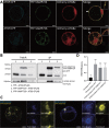
VAMP724 and VAMP726 are associated with ATG9 in Arabidopsis. (A) Subcellular localization analysis among ATG9, ATG8 and VAMP724 or VAMP726. The triple-constructs were transiently expressed together in Arabidopsis protoplasts and incubated for 12 h, followed by confocal imaging analysis. Arrows indicated examples of colocalization among the three fusions proteins with enlargement in the inset. Bar: 10 μm. (B) Immunoprecipitation (IP) assay shows association between ATG9 and VAMP724 or VAMP726. Arabidopsis protoplasts co-expressing ATG9-5FLAG with YFP (lane 1), YFP-VAMP724 (lane 2), or YFP-VAMP726 (lane 3) were subjected to protein extraction and IP with GFP-trap, followed by immunoblot analysis with indicated antibodies. Arrowhead indicates the ATG9 proteins immunoprecipitated by VAMP724 or VAMP726. (C) FRET analysis of the colocalized puncta between VAMP724 and ATG9 (left) or VAMP726 and ATG9 (right). The dashed oval represents the region for photobleaching. Bar: 10 μm. (D) Quantification of FRET efficiency using the acceptor photobleaching approach. 10 individual protoplasts were used for the statistical analysis. Error bars are the S.D. ****P < 0.0001, two-sided Student’s t-test.
Figure 8.
Figure 8.
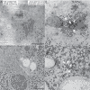
ATG9 resides on TGN and traffics together with VAMP726 to the forming autophagosome upon autophagy-induction. Ultrathin sections were prepared from high-pressure frozen/freeze-substituted root cells of YFP-VAMP726 ATG9-mCherry seedlings upon 6-h BTH treatment, followed by double immunogold labeling with anti-GFP (6-nm gold, indicated by arrows) and anti-RFP antibodies (10-nm gold, indicated by arrowheads). (A) ATG9 resides on the TGN (trans-Golgi network) labeled by VAMP726. (B) Enlargement of the indicated box in (A). (C) A membrane expanding phagophore. (D) Enlargement of the indicated box in (C), both anti-ATG9 and anti-VAMP726 antibodies labeled the edge of an expanding phagophore. Bar: 500 nm.

Similar articles

Cited by

References

    1. Xie Z, Klionsky DJ.. Autophagosome formation: core machinery and adaptations. Nat Cell Biol. 2007;9(10):1102–9. - PubMed
    1. Liu Y, Bassham DC.. Autophagy: pathways for self-eating in plant cells. Annu Rev Plant Biol. 2012;63:215–37. - PubMed
    1. Morishita H, Mizushima N.. Diverse cellular roles of autophagy. Annu Rev Cell Dev Biol. 2019;35:453–475. - PubMed
    1. Lipka V, Kwon C, Panstruga R.. SNARE-ware: the role of SNARE-domain proteins in plant biology. Annu Rev Cell Dev Biol. 2007;23:147–74. - PubMed
    1. Nair U, Jotwani A, Geng J, et al. SNARE proteins are required for macroautophagy. Cell. 2011;146(2):290–302. - PMC - PubMed

Publication types

MeSH terms

Substances

Grants and funding

This work was supported by grants from the National Natural Science Foundation of China (91854201), the Research Grants Council of Hong Kong (AoE/M-05/12, CUHK 14100818, 14101219, C4033-19E, C4002-17G, C4002-20W, C4002-21EF, C2009-19G, and R4005-18), and The Chinese University of Hong Kong (CUHK) Research Committee and CAS-Croucher Funding Scheme for Joint Laboratories to L.J; the National Natural Science Foundation of China (32061160467 and 31870171) and Fok Ying-Tong Education Foundation for Young Teachers in the Higher Education Institutions of China (171014) to C.G.

LinkOut - more resources