

Stimulation of the ventromedial prefrontal cortex blocks the return of subcortically mediated fear responses
- PMID: 36127327
- PMCID: PMC9489865
- DOI: 10.1038/s41398-022-02174-8
Stimulation of the ventromedial prefrontal cortex blocks the return of subcortically mediated fear responses
Abstract
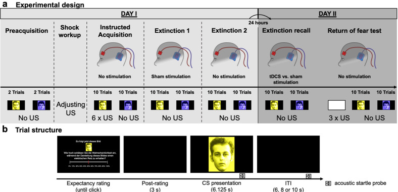
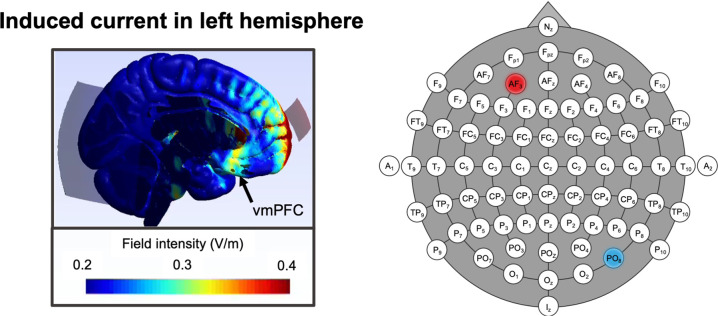
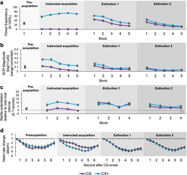
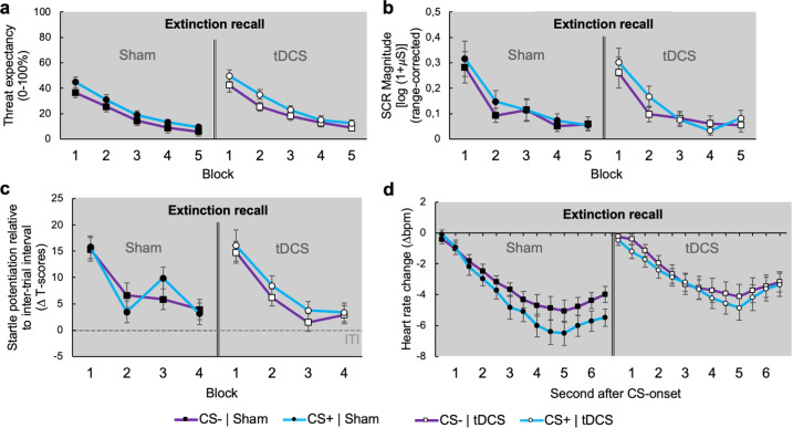
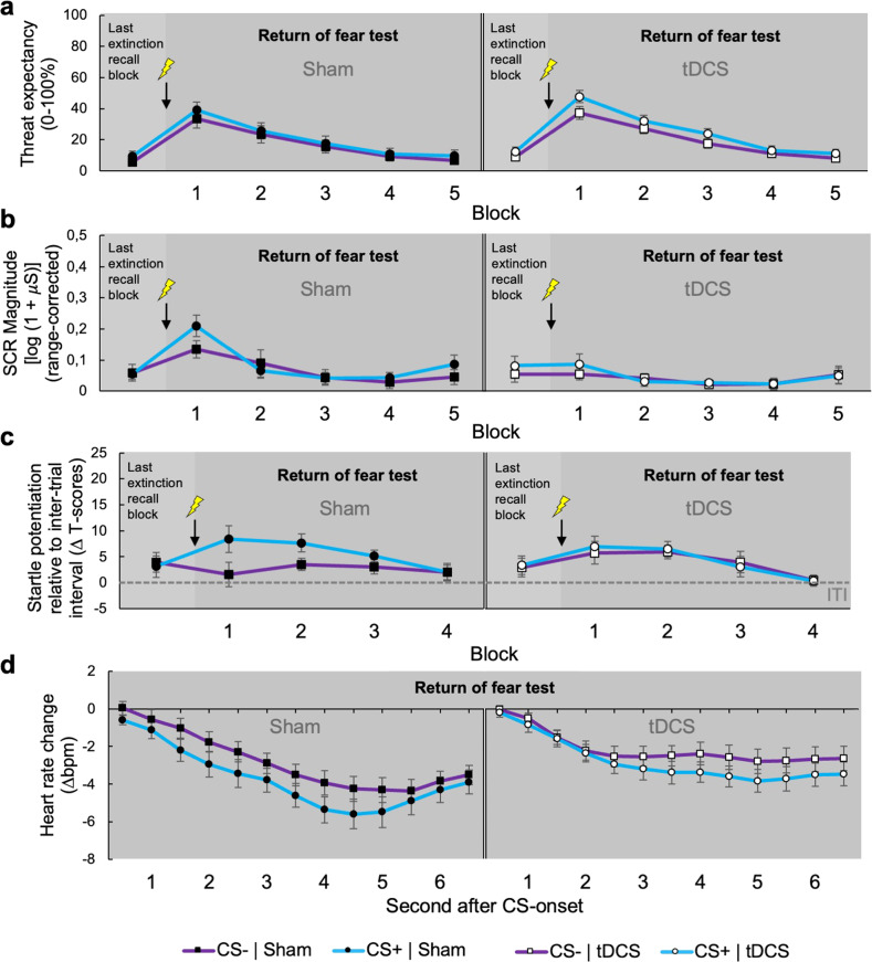
The ventromedial prefrontal cortex (vmPFC) mediates the inhibition of defensive responses upon encounters of cues, that had lost their attribute as a threat signal via previous extinction learning. Here, we investigated whether such fear extinction recall can be facilitated by anodal transcranial direct current stimulation (tDCS). Extinction recall was tested twenty-four hours after previously acquired fear was extinguished. Either anodal tDCS or sham stimulation targeting the vmPFC was applied during this test. After stimulation ceased, we examined return of fear after subjects had been re-exposed to aversive events. Fear was assessed by reports of threat expectancy and modulations of autonomic (skin conductance, heart rate) and protective reflex (startle potentiation) measures, the latter of which are mediated by subcortical defense circuits. While tDCS did not affect initial extinction recall, it abolished the return of startle potentiation and autonomic components of the fear response. Results suggest hierarchical multi-level vmPFC functions in human fear inhibition and indicate, that its stimulation might immunize against relapses into pathological subcortically mediated defensive activation.
© 2022. The Author(s).
Conflict of interest statement
The authors declare no competing interests.
Figures
Similar articles
-
Transcranial direct current stimulation may modulate extinction memory in posttraumatic stress disorder.Brain Behav. 2017 Apr 11;7(5):e00681. doi: 10.1002/brb3.681. eCollection 2017 May. Brain Behav. 2017. PMID: 28523223 Free PMC article.
-
Good moments to stimulate the brain - A randomized controlled double-blinded study on anodal transcranial direct current stimulation of the ventromedial prefrontal cortex on two different time points in a two-day fear conditioning paradigm.Behav Brain Res. 2024 Mar 5;460:114804. doi: 10.1016/j.bbr.2023.114804. Epub 2023 Dec 14. Behav Brain Res. 2024. PMID: 38103872 Clinical Trial.
-
Can Transcranial Direct Current Stimulation Augment Extinction of Conditioned Fear?Brain Stimul. 2016 Jul-Aug;9(4):529-36. doi: 10.1016/j.brs.2016.03.004. Epub 2016 Mar 9. Brain Stimul. 2016. PMID: 27037186 Free PMC article.
-
The effect of transcranial direct current and magnetic stimulation on fear extinction and return of fear: A meta-analysis and systematic review.J Affect Disord. 2024 Oct 1;362:263-286. doi: 10.1016/j.jad.2024.06.060. Epub 2024 Jun 21. J Affect Disord. 2024. PMID: 38908557 Review.
-
Prefrontal but not cerebellar tDCS attenuates renewal of extinguished conditioned eyeblink responses.Neurobiol Learn Mem. 2020 Apr;170:107137. doi: 10.1016/j.nlm.2019.107137. Epub 2019 Dec 12. Neurobiol Learn Mem. 2020. PMID: 31838223 Review.
Cited by
-
Effectiveness of repetitive transcranial magnetic stimulation for insomnia disorder on fear memory extinction: study protocol for a randomised controlled trial.Trials. 2024 Jun 19;25(1):396. doi: 10.1186/s13063-024-08198-3. Trials. 2024. PMID: 38898471 Free PMC article.
-
Frontopolar multifocal transcranial direct current stimulation reduces conditioned fear reactivity during extinction training: A pilot randomized controlled trial.Neurobiol Learn Mem. 2023 Nov;205:107825. doi: 10.1016/j.nlm.2023.107825. Epub 2023 Sep 10. Neurobiol Learn Mem. 2023. PMID: 37699439 Free PMC article. Clinical Trial.
References
Publication types
MeSH terms
LinkOut - more resources
Full Text Sources
