Lipopolysaccharide sensitizes the therapeutic response of breast cancer to IAP antagonist
- PMID: 36119107
- PMCID: PMC9471085
- DOI: 10.3389/fimmu.2022.906357
Lipopolysaccharide sensitizes the therapeutic response of breast cancer to IAP antagonist
Abstract
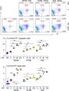
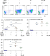
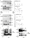
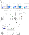
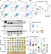
Inhibitor of apoptosis protein (IAP) is a class of E3 ubiquitin ligases functioning to support cancer survival and growth. Many small-molecule IAP antagonists have been developed, aiming to degrade IAP proteins to kill cancer. We have evaluated the effect of lipopolysaccharide (LPS), a component of the bacterial outer membrane, on IAP antagonists in treating breast cancer in a mouse model to guide future clinical trials. We show that LPS promotes IAP antagonist-induced regression of triple-negative breast cancer (TNBC) from MDA-MB-231 cells in immunodeficient mice. IAP antagonists such as SM-164, AT-406, and BV6, do not kill MDA-MB-231 cells alone, but allow LPS to induce cancer cell apoptosis rapidly. The apoptosis caused by LPS plus SM-164 is blocked by toll-like receptor 4 (TLR4) or MyD88 inhibitor, which inhibits LPS-induced TNFα production by the cancer cells. Consistent with this, MDA-MB-231 cell apoptosis induced by LPS plus SM-164 is also blocked by the TNF inhibitor. LPS alone does not kill MDA-MB-231 cells because it markedly increases the protein level of cIAP1/2, which is directly associated with and stabilized by MyD88, an adaptor protein of TLR4. ER+ MCF7 breast cancer cells expressing low levels of cIAP1/2 undergo apoptosis in response to SM-164 combined with TNFα but not with LPS. Furthermore, TNFα but not LPS alone inhibits MCF7 cell growth in vitro. Consistent with these, LPS combined with SM-164, but not either of them alone, causes regression of ER+ breast cancer from MCF7 cells in immunodeficient mice. In summary, LPS sensitizes the therapeutic response of both triple-negative and ER+ breast cancer to IAP antagonist therapy by inducing rapid apoptosis of the cancer cells through TLR4- and MyD88-mediated production of TNFα. We conclude that antibiotics that can reduce microbiota-derived LPS should not be used together with an IAP antagonist for cancer therapy.
Keywords: MyD88; TNF-α; Tolllike receptor 4; apoptosis; breast cancer; inhibitor of apoptosis protein; lipopolysaccharide.
Copyright © 2022 Liu, Yao, Chen, Lei, Duan and Yao.
Conflict of interest statement
The authors declare that the research was conducted in the absence of any commercial or financial relationships that could be construed as a potential conflict of interest.
Figures


Similar articles
-
Chimeric antigen receptor dendritic cells targeted delivery of a single tumoricidal factor for cancer immunotherapy.Cancer Immunol Immunother. 2024 Aug 6;73(10):203. doi: 10.1007/s00262-024-03788-1. Cancer Immunol Immunother. 2024. PMID: 39105847 Free PMC article.
-
Lipopolysaccharide inhibition of glucose production through the Toll-like receptor-4, myeloid differentiation factor 88, and nuclear factor kappa b pathway.Hepatology. 2009 Aug;50(2):592-600. doi: 10.1002/hep.22999. Hepatology. 2009. PMID: 19492426 Free PMC article.
-
A small molecule Smac-mimic compound induces apoptosis and sensitizes TRAIL- and etoposide-induced apoptosis in breast cancer cells.Oncogene. 2005 Nov 10;24(49):7381-8. doi: 10.1038/sj.onc.1208888. Oncogene. 2005. PMID: 16044155
-
IAP antagonists target cIAP1 to induce TNFalpha-dependent apoptosis.Cell. 2007 Nov 16;131(4):682-93. doi: 10.1016/j.cell.2007.10.037. Cell. 2007. PMID: 18022363
-
Toll-like receptor 4 prompts human breast cancer cells invasiveness via lipopolysaccharide stimulation and is overexpressed in patients with lymph node metastasis.PLoS One. 2014 Oct 9;9(10):e109980. doi: 10.1371/journal.pone.0109980. eCollection 2014. PLoS One. 2014. PMID: 25299052 Free PMC article.
Cited by
-
LINC00173 silence and estrone supply suppress ER+ breast cancer by estrogen receptor α degradation and LITAF activation.Cancer Sci. 2024 Jul;115(7):2318-2332. doi: 10.1111/cas.16201. Epub 2024 May 5. Cancer Sci. 2024. PMID: 38705575 Free PMC article.
-
Targeting Store-Operated Calcium Entry Regulates the Inflammation-Induced Proliferation and Migration of Breast Cancer Cells.Biomedicines. 2023 Jun 4;11(6):1637. doi: 10.3390/biomedicines11061637. Biomedicines. 2023. PMID: 37371732 Free PMC article.
-
MyD88 signaling pathways: role in breast cancer.Front Oncol. 2024 Jan 29;14:1336696. doi: 10.3389/fonc.2024.1336696. eCollection 2024. Front Oncol. 2024. PMID: 38347830 Free PMC article. Review.
-
Lipopolysaccharide promotes cancer cell migration and invasion through METTL3/PI3K/AKT signaling in human cholangiocarcinoma.Heliyon. 2024 Apr 16;10(8):e29683. doi: 10.1016/j.heliyon.2024.e29683. eCollection 2024 Apr 30. Heliyon. 2024. PMID: 38681552 Free PMC article.
-
Chimeric antigen receptor dendritic cells targeted delivery of a single tumoricidal factor for cancer immunotherapy.Cancer Immunol Immunother. 2024 Aug 6;73(10):203. doi: 10.1007/s00262-024-03788-1. Cancer Immunol Immunother. 2024. PMID: 39105847 Free PMC article.
References
-
- Clarke M, Collins R, Darby S, Davies C, Elphinstone P, Evans V, et al. . Effects of radiotherapy and of differences in the extent of surgery for early breast cancer on local recurrence and 15-year survival: an overview of the randomised trials. Lancet (2005) 366:2087–106. doi: 10.1016/S0140-6736(05)67887-7 - DOI - PubMed
Publication types
MeSH terms
Substances
Grants and funding
LinkOut - more resources
Full Text Sources
Research Materials
Miscellaneous

