Review
. 2022 Oct 15;400(10360):1363-1380.
doi: 10.1016/S0140-6736(22)01272-7. Epub 2022 Sep 15.

Amyotrophic lateral sclerosis

Affiliations
Review

Amyotrophic lateral sclerosis

Eva L Feldman et al. Lancet. .

Abstract

Amyotrophic lateral sclerosis is a fatal CNS neurodegenerative disease. Despite intensive research, current management of amyotrophic lateral sclerosis remains suboptimal from diagnosis to prognosis. Recognition of the phenotypic heterogeneity of amyotrophic lateral sclerosis, global CNS dysfunction, genetic architecture, and development of novel diagnostic criteria is clarifying the spectrum of clinical presentation and facilitating diagnosis. Insights into the pathophysiology of amyotrophic lateral sclerosis, identification of disease biomarkers and modifiable risks, along with new predictive models, scales, and scoring systems, and a clinical trial pipeline of mechanism-based therapies, are changing the prognostic landscape. Although most recent advances have yet to translate into patient benefit, the idea of amyotrophic lateral sclerosis as a complex syndrome is already having tangible effects in the clinic. This Seminar will outline these insights and discuss the status of the management of amyotrophic lateral sclerosis for the general neurologist, along with future prospects that could improve care and outcomes for patients with amyotrophic lateral sclerosis.


Conflict of interest statement

Declaration of interests ELF and SAG have a patent issued (US20200253977A1). SAG reports personal fees from Biogen, ITF Pharma, and Watermark, outside the submitted work. SP reports grants from the German Neuromuscular Society, the German-Israeli Foundation for Scientific Research and Development (GIF), and personal fees from Cytokinetics, Desitin Pharma, Italfarmaco, Biogen, Roche, and Zambon outside the submitted work. PJS reports consultancy and advisory board membership with Biogen, Benevolent AI, QurALIS, Quell, and Aclipse Therapeutics, outside the submitted work. GS reports personal fees from Mitsubishi Tanabe Pharma Corporation, Cyberdyne, Biogen Japan, Takeda Pharmaceutical, Nihon Pharmaceutical, and Teijin Pharma, outside the submitted work. LM and MGS declare no competing interests.

Figures

Figure 1.
Figure 1.. ALS phenotypic variation and spectrum with FTD.
(A) Schematic showing upper motor neurons (UMN; blue), which relay signals from the motor cortex to the lower motor neurons (LMN; yellow, i.e., cranial motor nerve nuclei in the brainstem and anterior horn cells in the spinal cord), which relay signals to the muscles. Motor neurons connecting within the brain stem innervate, among other muscles, cranial muscles. Initial UMN and LMN degeneration in the brain stem are linked to bulbar onset ALS. Motor neurons connecting within the cervical region of the spinal cord innervate, among other muscles, upper limb and respiratory muscles. Motor neurons connecting within the thoracic and lumbar regions of the spinal cord innervate, among other muscles, accessory respiratory, abdominal, and lower limb muscles. Initial UMN and LMN degeneration in the cervical and lumbar regions are linked to spinal onset ALS. (B) ALS patients can present with signs of UMN (blue), LMN (yellow), and combined UMN + LMN (green) dysfunction. Most common ALS phenotypic presentations are bulbar and classical spinal limb onset (cervical, lumbar). Less common ALS phenotypic presentations are flail leg, pyramidal, flail arm, primary lateral sclerosis (PLS), progressive muscular atrophy (PMA), respiratory onset, and hemiplegic. Proportion of various ALS phenotypes shown in the figure as the percentage (%) of a total representative ALS population.(14, 17) Pyramidal is predominantly UMN, as shown in the figure, but still exhibits some LMN signs, differentiating it from PLS. See Appendix Table 1 for more information. (C) ALS occurs on a continuum with FTD. ALS is on one end of the spectrum and presents with pure motor signs from UMN + LMN neurodegeneration (green, spinal cord and motor cortex degeneration). FTD is on the other end of the spectrum and presents with behavioral and/or cognitive deficits from frontotemporal neurodegeneration (purple frontotemporal lobe degeneration). After pure ALS are ALS patients not meeting FTD criteria, defined as ALS cognitive impairment (ALSci), ALS behavioral impairment (ALSbi), and ALS cognitive and behavioral impairment (ALScbi) (green, spinal cord and motor cortex degeneration; small purple sphere of frontotemporal lobe degeneration). Next are ALS patients meeting FTD criteria, defined as ALS-FTD (green, spinal cord and motor cortex degeneration; purple frontotemporal lobe degeneration). Patients on the remainder of the continuum have FTD but do not meet the criteria for ALS. Patients still exhibiting evidence of motor neuron disease (MND) with FTD are defined as MND-FTD (dark grey, spinal cord and motor cortex degeneration; purple frontotemporal lobe degeneration) and patients with no MND signs have FTD (purple, frontotemporal lobe degeneration).
Figure 2.
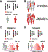
Figure 2.. ALS genetic architecture.
ALS genetics is characterized by monogenic, oligogenic, and polygenic risk; image featuring only three representative chromosomes (within each panel, chromosomes on the left for healthy person, on the right for person with ALS). (A) Monogenic inheritance in ALS, characterized by inheritance of a single gene. (B) ALS genes are not fully penetrant and pathogenicity of certain variants is uncertain. Left: In a population of gene carriers, low penetrance variants lead to a low frequency of ALS onset (red figures). Right: In a population of gene carriers, high penetrance variants lead to a high frequency of ALS onset (red figures). (C) Oligogenic inheritance in ALS, characterized by inheritance of several genes (four shown in the figure). (D) Polygenic inheritance in ALS, characterized by inheritance of many genes (nine shown in the figure). Created with BioRender.com. Adapted, with permission, from Goutman et al. The Lancet Neurology, 2022.
Figure 3.
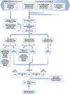
Figure 3.. ALS differential diagnosis.
Differential diagnosis, represented here by a flowchart for the classical process using symptoms and signs, is central to the diagnostic process in ALS. At minimum, individuals suspected of ALS will undergo physical and neurological exams, electrodiagnostic assessment, MRI of involved regions, and relevant serological testing. This figure is based on a summary of potential differential diagnoses for diseases more common or as common as ALS is outlined in Appendix Table 3. Overlap of known ALS genes with other diseases and syndromes also occurs and is outlined in Appendix Table 4. CFS, cramp-fasciculation syndrome; CIDP, chronic inflammatory demyelinating polyneuropathy; HSP, hereditary spastic paraparesis; IBM, inclusion body myositis; LMN, lower motor neuron; MMN, multifocal motor neuropathy; MG, myasthenia gravis; PPMS primary progressive multiple sclerosis; rEEC, revise El Escorial criteria; SBMA, spinobulbar muscular atrophy; UMN, upper motor neuron. *Several potential differential diagnoses present with proximal weakness and should be considered along with flail arm ALS, which also presents with proximal greater than distal upper extremity weakness. Thus, check for increased proximal reflexes on exam and neurogenic motor unit action potentials on electromyography.
Figure 4.
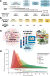
Figure 4.. ALS risk and prognosis.
(A) King’s staging with four stages indicated (1, 2A/B, 3, 4A/B; blue); time to progress to stages and median survival at each stage (in months) are also annotated. (B) ALS-MiToS staging with six stages indicated (0, 1, 2, 3, 4, 5; orange); staging based on four functional domains from the ALSFRS-R: (i) movement (walking/self-care; ALSFRS-R question 6 or 8); (ii) swallowing (ALSFRS-R question 3); (iii) communicating (ALSFRS-R questions 1 and 4), and (iv) breathing (ALSFRS-R question 10 or 12). Intensifying color indicates progression along stages for both King’s and ALS-MiToS. (C) Schematic overview of factors that affect ALS risk (onset) and prognosis, which include clinical and demographic features (e.g., age at onset, segment onset, ALSFRS-R progression rate, forced vital capacity, FTD), genetic architecture (e.g., rapidly progressive SOD1A5V, slowly progressive DCTN1 mutations), and exposome (e.g., environmental exposures). (D) ENCALS prediction model of ALS prognosis, represented, with permission, from Westeneng, The Lancet Neurology, 2018.(70) The model leverages 8 clinical predictors to the composite endpoint (survival without tracheostomy or non-invasive ventilation >23 hours per day): age at onset, time to diagnosis, ALSFRS-R progression rate, forced vital capacity, bulbar onset, definite ALS by revised El Escorial criteria, FTD, and C9orf72 repeat expansion. Top: The model defines five survival groups: very short (red; predicted median survival [MS] 17·7 months), short (orange; predicted MS 25·3 months), intermediate (light orange; predicted MS 32·2 months), long (light green; predicted MS 43·7 months), and very long (predicted MS green; 91·0 months). The dashed black line represents MS without employing the ENCALS prediction model, which is overly optimistic for ALS patients classified to the very short and short survival groups, i.e., they end up with less time, and overly pessimistic for patients classified to long and very long groups, i.e., they end up with more time. Bottom: Horizontal bars have dots to represent median times to composite outcome, thick lines to represent probability interquartile range, thin lines to represent 10 to 90% probability intervals to composite outcome. Created, in part, with BioRender.com.

Similar articles

Cited by

References

    1. Ryan M, Heverin M, McLaughlin RL, Hardiman O. Lifetime Risk and Heritability of Amyotrophic Lateral Sclerosis. JAMA Neurol. 2019;76(11):1367–74. - PMC - PubMed
    1. Goutman SA, Hardiman O, Al-Chalabi A, Chió A, Savelieff MG, Kiernan MC, et al. Recent advances in the diagnosis and prognosis of amyotrophic lateral sclerosis. Lancet Neurol. 2022. - PMC - PubMed
    1. Marin B, Fontana A, Arcuti S, Copetti M, Boumédiene F, Couratier P, et al. Age-specific ALS incidence: a dose-response meta-analysis. Eur J Epidemiol. 2018;33(7):621–34. - PubMed
    1. Mehta P, Kaye W, Raymond J, Punjani R, Larson T, Cohen J, et al. Prevalence of Amyotrophic Lateral Sclerosis - United States, 2015. MMWR Morb Mortal Wkly Rep. 2018;67(46):1285–9. - PMC - PubMed
    1. Luna J, Diagana M, Ait Aissa L, Tazir M, Ali Pacha L, Kacem I, et al. Clinical features and prognosis of amyotrophic lateral sclerosis in Africa: the TROPALS study. J Neurol Neurosurg Psychiatry. 2019;90(1):20–9. - PubMed