Mechanobiological responses of astrocytes in optic nerve head due to biaxial stretch
- PMID: 36114477
- PMCID: PMC9482189
- DOI: 10.1186/s12886-022-02592-8
Mechanobiological responses of astrocytes in optic nerve head due to biaxial stretch
Abstract
Background: Elevated intraocular pressure (IOP) is the main risk factor for glaucoma, which might cause the activation of astrocytes in optic nerve head. To determine the effect of mechanical stretch on the astrocytes, we investigated the changes in cell phenotype, proteins of interest and signaling pathways under biaxial stretch.
Method: The cultured astrocytes in rat optic nerve head were stretched biaxially by 10 and 17% for 24 h, respectively. Then, we detected the morphology, proliferation and apoptosis of the stretched cells, and performed proteomics analysis. Protein expression was analyzed by Isobaric tags for relative and absolute quantification (iTRAQ) mass spectrometry. Proteins of interest and signaling pathways were screened using Gene Ontology enrichment analysis and pathway enrichment analysis, and the results were verified by western blot and the gene-chip data from Gene Expression Omnibus (GEO) database.
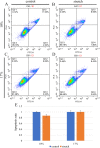
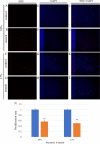
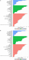
Result: The results showed that rearrangement of the actin cytoskeleton in response to stimulation by mechanical stress and proliferation rate of astrocytes decreased under 10 and 17% stretch condition, while there was no significant difference on the apoptosis rate of astrocytes in both groups. In the iTRAQ quantitative experiment, there were 141 differential proteins in the 10% stretch group and 140 differential proteins in the 17% stretch group. These proteins include low-density lipoprotein receptor-related protein (LRP6), caspase recruitment domain family, member 10 (CARD10), thrombospondin 1 (THBS1) and tetraspanin (CD81). The western blot results of LRP6, THBS1 and CD81 were consistent with that of iTRAQ experiment. ANTXR2 and CARD10 were both differentially expressed in the mass spectrometry results and GEO database. We also screened out the signaling pathways associated with astrocyte activation, including Wnt/β-catenin pathway, NF-κB signaling pathway, PI3K-Akt signaling pathway, MAPK signaling pathway, Jak-STAT signaling pathway, ECM-receptor interaction, and transforming growth factor-β (TGF-β) signaling pathway.
Conclusion: Mechanical stimulation can induce changes in cell phenotype, some proteins and signaling pathways, which might be associated with astrocyte activation. These proteins and signaling pathways may help us have a better understanding on the activation of astrocytes and the role astrocyte activation played in glaucomatous optic neuropathy.
Keywords: Astrocytes; Biaxial stretch; Mechanobiological responses; Optic nerve head; Proteomics.
© 2022. The Author(s).
Conflict of interest statement
The authors declare that they have no competing interests.
Figures


Similar articles
-
Transforming growth factor-β2 increases extracellular matrix proteins in optic nerve head cells via activation of the Smad signaling pathway.Mol Vis. 2011;17:1745-58. Epub 2011 Jun 28. Mol Vis. 2011. PMID: 21738403 Free PMC article.
-
Proteomics analyses of human optic nerve head astrocytes following biomechanical strain.Mol Cell Proteomics. 2012 Feb;11(2):M111.012302. doi: 10.1074/mcp.M111.012302. Epub 2011 Nov 29. Mol Cell Proteomics. 2012. PMID: 22126795 Free PMC article.
-
Regional Gene Expression in the Retina, Optic Nerve Head, and Optic Nerve of Mice with Optic Nerve Crush and Experimental Glaucoma.Int J Mol Sci. 2023 Sep 6;24(18):13719. doi: 10.3390/ijms241813719. Int J Mol Sci. 2023. PMID: 37762022 Free PMC article.
-
Integrins in trabecular meshwork and optic nerve head: possible association with the pathogenesis of glaucoma.Biomed Res Int. 2013;2013:202905. doi: 10.1155/2013/202905. Epub 2013 Mar 18. Biomed Res Int. 2013. PMID: 23586020 Free PMC article. Review.
-
The role of astrocytes in optic nerve head fibrosis in glaucoma.Exp Eye Res. 2016 Jan;142:49-55. doi: 10.1016/j.exer.2015.08.014. Epub 2015 Aug 29. Exp Eye Res. 2016. PMID: 26321510 Review.
Cited by
-
Morphological comparison of astrocytes in the lamina cribrosa and glial lamina.bioRxiv [Preprint]. 2024 Sep 10:2024.09.07.610493. doi: 10.1101/2024.09.07.610493. bioRxiv. 2024. PMID: 39314351 Free PMC article. Preprint.
-
Meconopsis quintuplinervia Regel Improves Cutibacterium acnes-Induced Inflammatory Responses in a Mouse Ear Edema Model and Suppresses Pro-Inflammatory Chemokine Production via the MAPK and NF-κB Pathways in RAW264.7 Cells.Ann Dermatol. 2023 Dec;35(6):408-416. doi: 10.5021/ad.22.206. Ann Dermatol. 2023. PMID: 38086354 Free PMC article.
-
The effect of the mechanodynamic lung environment on fibroblast phenotype via the Flexcell.BMC Pulm Med. 2024 Jul 27;24(1):362. doi: 10.1186/s12890-024-03167-7. BMC Pulm Med. 2024. PMID: 39068387 Free PMC article. Review.
-
Engineered cell culture microenvironments for mechanobiology studies of brain neural cells.Front Bioeng Biotechnol. 2022 Dec 14;10:1096054. doi: 10.3389/fbioe.2022.1096054. eCollection 2022. Front Bioeng Biotechnol. 2022. PMID: 36588937 Free PMC article. Review.
-
Long-term menopause exacerbates vaginal wall support injury in ovariectomized rats by regulating amino acid synthesis and glycerophospholipid metabolism.Front Endocrinol (Lausanne). 2023 Jun 22;14:1119599. doi: 10.3389/fendo.2023.1119599. eCollection 2023. Front Endocrinol (Lausanne). 2023. PMID: 37424873 Free PMC article.
References
-
- Tehrani S, Davis L, Cepurna WO, Choe TE, Lozano DC, Monfared A, et al. Astrocyte structural and molecular response to elevated intraocular pressure occurs rapidly and precedes axonal tubulin rearrangement within the optic nerve head in a rat model. PLoS ONE. 2016;11:e0167364. doi: 10.1371/journal.pone.0167364. - DOI - PMC - PubMed
MeSH terms
Substances
LinkOut - more resources
Full Text Sources
Medical
Miscellaneous

