Comparative effects of free doxorubicin, liposome encapsulated doxorubicin and liposome co-encapsulated alendronate and doxorubicin (PLAD) on the tumor immunologic milieu in a mouse fibrosarcoma model
- PMID: 36105861
- PMCID: PMC9461478
- DOI: 10.7150/ntno.75045
Comparative effects of free doxorubicin, liposome encapsulated doxorubicin and liposome co-encapsulated alendronate and doxorubicin (PLAD) on the tumor immunologic milieu in a mouse fibrosarcoma model
Abstract
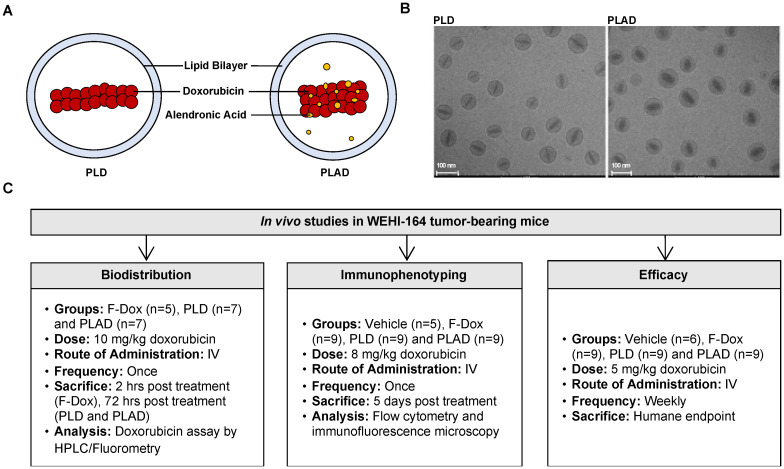
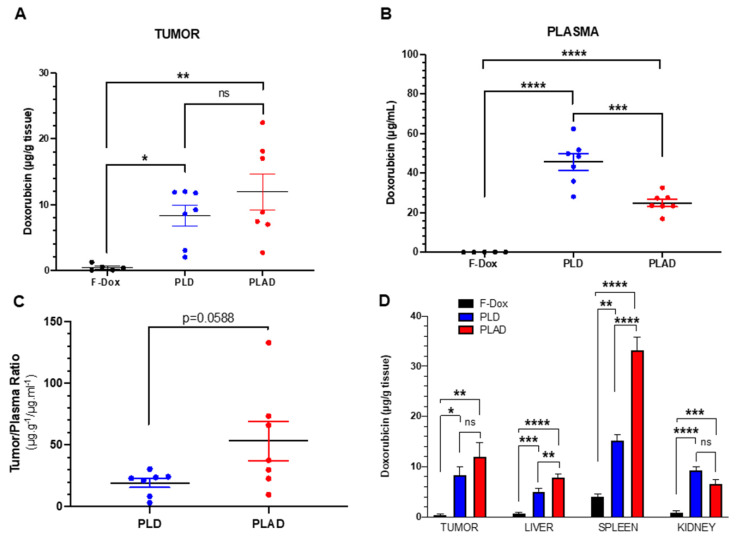
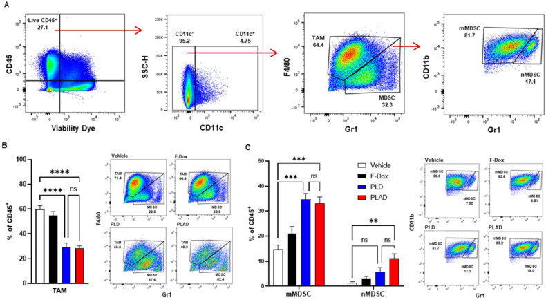
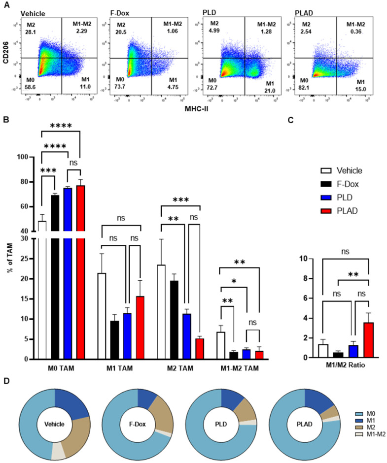
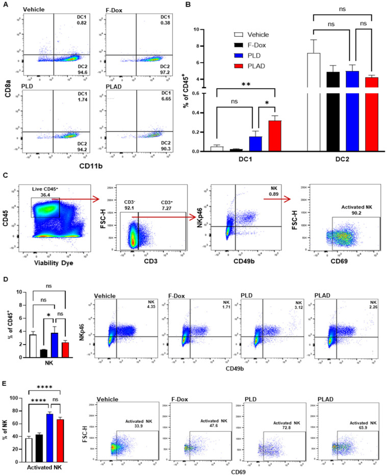
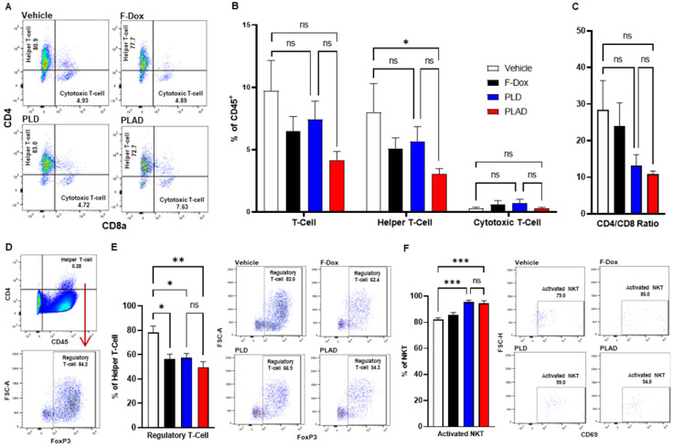
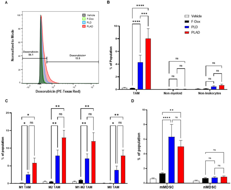
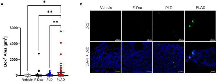
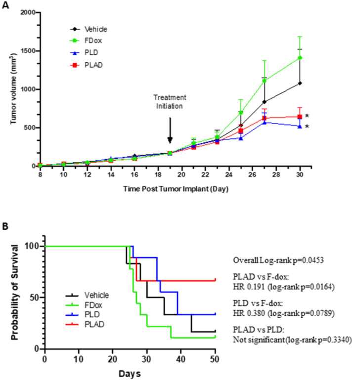
Background: We have previously shown that alendronate, an amino-bisphosphonate, when reformulated in liposomes, can significantly enhance the efficacy of cytotoxic chemotherapies and help remodel the immunosuppressive tumor microenvironment towards an immune-permissive milieu resulting in increased anticancer efficacy. In addition, we have previously shown that the strong metal-chelating properties of alendronate can be exploited for nuclear imaging of liposomal biodistribution. To further improve anticancer efficacy, a pegylated liposome formulation co-encapsulating alendronate and doxorubicin (PLAD) has been developed. In this study, we examined the effects of PLAD on the tumor immunologic milieu in a mouse fibrosarcoma model in which the tumor microenvironment is heavily infiltrated with tumor-associated macrophages (TAM) that are associated with poor prognosis and treatment resistance. Methods: Doxorubicin biodistribution, characterization of the tumor immunologic milieu, cellular doxorubicin uptake, and tumor growth studies were performed in Balb/c mice bearing subcutaneously implanted WEHI-164 fibrosarcoma cells treated intravenously with PLAD, pegylated liposomal doxorubicin (PLD), free doxorubicin, or vehicle. Results: PLAD delivery resulted in a high level of tumor doxorubicin that was 20 to 30-fold greater than in free doxorubicin treated mice, and non-significantly higher than in PLD treated mice. PLAD also resulted in increased uptake in spleen and slightly lower plasma levels as compared to PLD. Importantly, our results showed that PLAD, and to a lesser extent PLD, shifted cellular drug uptake to TAM and to monocytic myeloid-derived suppressor cells (MDSC), while there was no drug uptake in neutrophilic MDSC or lymphoid cells. Free doxorubicin cellular drug uptake was below detectable levels. PLAD, and to a lesser extent PLD, also induced significant changes in number and functionality of tumor-infiltrating TAM, MDSC, Treg, NKT, and NK cells that are consistent with enhanced antitumor immune responses in the tumor microenvironment. In contrast, free doxorubicin induced moderate changes in the tumor microenvironment that could promote (decreased Treg) or be detrimental to antitumor immune responses (decreased M1 TAM and NK cells). These immune modulatory effects are reflected in the therapeutic study which showed that PLAD and PLD inhibited tumor growth and significantly prolonged survival, while free doxorubicin showed little or no anticancer activity. Conclusion: We show that liposomal delivery of doxorubicin not only alters pharmacokinetics, but also dramatically changes the immune modulatory activity of the drug cargo. In addition, our data support that the PLAD nanotheranostic platform further enhances some immune changes that may act in synergy with its cytotoxic chemotherapy effects.
Keywords: alendronate; bisphosphonate; chemotherapy; doxorubicin; fibrosarcoma; immunotherapy; nanomedicine; tumor-associated macrophages.
© The author(s).
Conflict of interest statement
Competing Interests: The authors have declared that no competing interest exists.
Figures
Similar articles
-
Harnessing Nanomedicine to Potentiate the Chemo-Immunotherapeutic Effects of Doxorubicin and Alendronate Co-Encapsulated in Pegylated Liposomes.Pharmaceutics. 2023 Nov 9;15(11):2606. doi: 10.3390/pharmaceutics15112606. Pharmaceutics. 2023. PMID: 38004584 Free PMC article.
-
Liposome co-encapsulation of anti-cancer agents for pharmacological optimization of nanomedicine-based combination chemotherapy.Cancer Drug Resist. 2021 Jun 19;4(2):463-484. doi: 10.20517/cdr.2020.87. eCollection 2021. Cancer Drug Resist. 2021. PMID: 35582027 Free PMC article.
-
Coencapsulation of alendronate and doxorubicin in pegylated liposomes: a novel formulation for chemoimmunotherapy of cancer.J Drug Target. 2016 Nov;24(9):878-889. doi: 10.1080/1061186X.2016.1191081. Epub 2016 Jun 6. J Drug Target. 2016. PMID: 27187807
-
New insights and evolving role of pegylated liposomal doxorubicin in cancer therapy.Drug Resist Updat. 2016 Nov;29:90-106. doi: 10.1016/j.drup.2016.10.003. Epub 2016 Oct 29. Drug Resist Updat. 2016. PMID: 27912846 Review.
-
Clinical pharmacology of liposomal anthracyclines: focus on pegylated liposomal Doxorubicin.Clin Lymphoma Myeloma. 2008 Feb;8(1):21-32. doi: 10.3816/clm.2008.n.001. Clin Lymphoma Myeloma. 2008. PMID: 18501085 Review.
Cited by
-
Research progress on the mechanism of curcumin in cerebral ischemia/reperfusion injury: a narrative review.Apoptosis. 2023 Oct;28(9-10):1285-1303. doi: 10.1007/s10495-023-01869-7. Epub 2023 Jun 26. Apoptosis. 2023. PMID: 37358747 Review.
-
Harnessing Nanomedicine to Potentiate the Chemo-Immunotherapeutic Effects of Doxorubicin and Alendronate Co-Encapsulated in Pegylated Liposomes.Pharmaceutics. 2023 Nov 9;15(11):2606. doi: 10.3390/pharmaceutics15112606. Pharmaceutics. 2023. PMID: 38004584 Free PMC article.
-
Immune Implications of Cholesterol-Containing Lipid Nanoparticles.ACS Nano. 2024 Oct 22;18(42):28480-28501. doi: 10.1021/acsnano.4c06369. Epub 2024 Oct 10. ACS Nano. 2024. PMID: 39388645 Review.
-
A landscape of recent advances in lipid nanoparticles and their translational potential for the treatment of solid tumors.Bioeng Transl Med. 2023 Nov 9;9(2):e10601. doi: 10.1002/btm2.10601. eCollection 2024 Mar. Bioeng Transl Med. 2023. PMID: 38435821 Free PMC article. Review.
-
Overcoming neutrophil-induced immunosuppression in postoperative cancer therapy: Combined sialic acid-modified liposomes with scaffold-based vaccines.Asian J Pharm Sci. 2024 Apr;19(2):100906. doi: 10.1016/j.ajps.2024.100906. Epub 2024 Mar 16. Asian J Pharm Sci. 2024. PMID: 38595333 Free PMC article.
References
Publication types
MeSH terms
Substances
LinkOut - more resources
Full Text Sources