Review
. 2022 Oct 12;96(19):e0129722.
doi: 10.1128/jvi.01297-22. Epub 2022 Sep 14.

An Unexpected Encounter: Respiratory Syncytial Virus Nonstructural Protein 1 Interacts with Mediator Subunit MED25

Affiliations
Review

An Unexpected Encounter: Respiratory Syncytial Virus Nonstructural Protein 1 Interacts with Mediator Subunit MED25

Tessa Van Royen et al. J Virol. .

Abstract

Human respiratory syncytial virus (RSV) is the leading cause of severe acute lower respiratory tract infections in infants worldwide. Nonstructural protein NS1 of RSV modulates the host innate immune response by acting as an antagonist of type I and type III interferon (IFN) production and signaling in multiple ways. Likely, NS1 performs this function by interacting with different host proteins. In order to obtain a comprehensive overview of the NS1 interaction partners, we performed three complementary protein-protein interaction screens, i.e., BioID, MAPPIT, and KISS. To closely mimic a natural infection, the BioID proximity screen was performed using a recombinant RSV in which the NS1 protein is fused to a biotin ligase. Remarkably, MED25, a subunit of the Mediator complex, was identified in all three performed screening methods as a potential NS1-interacting protein. We confirmed the interaction between MED25 and RSV NS1 by coimmunoprecipitation, not only upon overexpression of NS1 but also with endogenous NS1 during RSV infection. We also demonstrate that the replication of RSV can be enhanced in MED25 knockout A549 cells, suggesting a potential antiviral role of MED25 during RSV infection. Mediator subunits function as transcriptional coactivators and are involved in transcriptional regulation of their target genes. Therefore, the interaction between RSV NS1 and cellular MED25 might be beneficial for RSV during infection by affecting host transcription and the host immune response to infection. IMPORTANCE Innate immune responses, including the production of type I and III interferons, play a crucial role in the first line of defense against RSV infection. However, only a poor induction of type I IFNs is observed during RSV infection, suggesting that RSV has evolved mechanisms to prevent type I IFN expression by the infected host cell. A unique RSV protein, NS1, is largely responsible for this effect, probably through interaction with multiple host proteins. A better understanding of the interactions that occur between RSV NS1 and host proteins may help to identify targets for an effective antiviral therapy. We addressed this question by performing three complementary protein-protein interaction screens and identified MED25 as an RSV NS1-interacting protein. We propose a role in innate anti-RSV defense for this Mediator complex subunit.

Keywords: MED25; interferon; nonstructural protein; protein-protein interactions; respiratory syncytial virus.


Conflict of interest statement

The authors declare no conflict of interest.

Figures
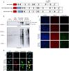
FIG 1
FIG 1
Characterization of recombinant RSVs expressing NS1-BirA* or mKate2-BirA*. (A) Genome organization (antigenomic cDNA) of recombinant parental RSV mKate2 and RSV expressing NS1-BirA* or mKate2-BirA*. (B) Immunoblot showing expression of NS1-BirA*-Flag and mKate2-BirA*-Flag fusion proteins (upper panel) and protein biotinylation upon expression of the fusion proteins (lower panel). A549 cells were infected with the recombinant BirA*-expressing RSVs (MOI, 1) or the control RSV mKate2 or were mock infected for 4 h, followed by incubation of the cells with 50 μM biotin for an additional 8, 16, or 24 h. Cell lysates were analyzed by Western blotting with an anti-Flag antibody or streptavidin. (C) Immunofluorescence images after infection of A549 cells with RSV NS1-BirA* and mKate2-BirA* (MOI, 1) for 4 h, followed by incubation with biotin (50 μM) for an additional 16 h. Cells were fixed and stained with streptavidin-AF488 and Hoechst stain. Scale bar, 0.1 mm. (D) A549 cells were infected with the indicated recombinant BirA*-expressing RSVs at an MOI of 1 or were mock infected for 4 h, followed by incubation of the cells with 50 μM biotin for an additional 16 h. The cells were then fixed and stained with streptavidin-AF488 and DAPI. The cells were imaged by confocal immunofluorescence microscopy. Scale bar, 10 μm.
FIG 2
FIG 2
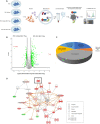
BioID screen with recombinant NS1-BirA*- and mKate2-BirA*-expressing RSV. (A) Schematic representation of the setup of the BioID screen with recombinant RSV NS1-BirA*-Flag and RSV mKate2-BirA*-Flag. A549 cells were infected (MOI, 1) for 4 h, followed by the addition of biotin (50 μM) for 16 h to biotinylate proteins in the proximity of NS1 and mKate2, respectively. After cell lysis (performed at 20 hpi), the biotinylated proteins were enriched on streptavidin-conjugated beads. The streptavidin-purified proteins were digested by trypsin on the beads, and the peptides were identified and quantified by LC-MS/MS analysis. The figure was created with BioRender software. (B) Volcano plot demonstrating fold change enrichment of host proteins in NS1-BirA* RSV-infected cells compared to mKate2-BirA* RSV-infected cells. Significantly enriched candidate interactors are depicted in green, blue (MED25), or red (NS1 and mKate2), and other identified proteins are depicted in gray (not significant). (C) Bioinformatics analysis of proteins that were identified in the BioID screen. All identified proteins were uploaded in the IPA software, with a filtering for significantly enriched proteins as indicated in panel B. An enrichment analysis was performed by IPA to assign molecular and cellular functions to the proteins enriched in the RSV NS1-BirA*-Flag-infected cells. (D) IPA software was used to explore the connections between proteins identified in the BioID screen in RSV NS1-BirA*-Flag-infected cells. Red and green nodes indicate proteins that are, respectively, significantly increased and decreased in abundance in RSV NS1-BirA*-Flag-infected cells compared to RSV mKate2-BirA*-Flag-infected cells, and the color intensity corresponds to the degree of abundance. White nodes represent proteins identified through the Ingenuity Pathways Knowledge Base. The biological relationship between two nodes is represented by a line. Solid and dashed lines indicate direct and indirect molecular relationships, respectively.
FIG 3
FIG 3
MAPPIT and KISS identify MED25 as a candidate NS1-interacting protein. (A) Results of the MAPPIT primary screening. A library covering 14,817 open reading frame (ORF) preys was screened with the NS1 bait chimera, with each ORF yielding quadruplicate samples for both the stimulated (+Epo) and unstimulated (−Epo) conditions. The plot represents the log-transformed q value of the ratio (stimulated versus unstimulated samples) of normalized MAPPIT luciferase activity versus the ratio (stimulated versus unstimulated samples) of the median value of fluorescence particle counts. (B) Results of the KISS primary screening. Each prey is transfected 4-fold with either the specific bait (NS1) or no bait (TYK2 only) as a control. The plot represents the log-transformed q value of the ratio (specific bait versus TYK2 only) of normalized KISS luciferase activity versus the ratio (specific bait versus TYK2 only) of the median value of fluorescence particle counts. For both panels A and B, a threshold q value of <0.35 and a particle count of >2 were applied, as these result in a high specificity (low false-positive rate). (C and D) Plots showing, respectively, the MAPPIT and KISS retests of 96 proteins of interest (list available in Table S4 in the supplemental material). The ORF preys were reevaluated by testing their interaction with either NS1 or a negative control bait. For MAPPIT, the fold induction of the average luciferase activity of triplicate EPO-stimulated versus unstimulated samples for either bait is shown. For KISS, the average luciferase activity for interaction with either bait is shown. A set of previously defined criteria for sensitive and specific MAPPIT and KISS analysis is applied (67): signals were scored negative (−; gray) or positive (+). Positive signals were further categorized as corresponding with an aspecific interaction (red) where the ORF prey binds to a component of the control bait rather than the NS1 bait or an NS1-specific interaction (green).
FIG 4
FIG 4
Venn diagram depicting the overlap of NS1 candidate interactors between the BioID, MAPPIT, and KISS screening methods. The BioID data set (significantly positively enriched proteins in RSV NS1-BirA*-Flag-infected samples relative to RSV mKate2-BirA*-Flag-infected samples), the MAPPIT and KISS data sets (filtered data set [q value of <0.35, particle counts of >2, removal of known aspecific binders] after primary screen against the 15,000 ORF prey collection) were imported into the jvenn online tool to identify proteins in common.
FIG 5
FIG 5
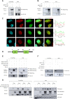
RSV NS1 binds to MED25. (A) Anti-Flag pulldown shows coimmunoprecipitation of NS1-HA with MED25-Flag but not with empty vector (EV) control after transient overexpression in HEK293T cells. Blots were analyzed with anti-Flag and anti-HA antibodies. (B) Anti-HA pulldown shows coimmunoprecipitation of MED25-Flag (left panel, lane 3) and endogenous MED25 (left panel, lane 2) with NS1-HA but not with EV control (left panel, lane 1). Blots were analyzed with anti-MED25 and anti-HA antibodies. (C) Representative confocal micrographs of HeLa cells that were transfected with NS1-GFP. Cells were fixed and stained with anti-MED25 antibody (red) or DAPI (blue). Scale bar, 10 μm. The three rows represent three different confocal images. The right panels show the intensity profile analysis along the white line (using ZEN 3.4 Blue software) to display the distribution of the red (MED25) and green (NS1-GFP) fluorophore signals. (D) Schematic representation of the MED25 domains. (E) Anti-Flag pulldown shows coimmunoprecipitation of NS1-HA with full-length MED25 (left panel, lane 2) and MED25 ACID (left panel, lane 3) but not with MED25 VWA (left panel, lane 4) or with EV (left panel, lane 1). Blots were analyzed with anti-Flag and anti-HA antibodies. (F) Anti-HA pulldown shows coimmunoprecipitation of MED25-Flag with NS1-HA (left panel, lane 2) but not with NS1 Δα3-HA (left panel, lane 3) or with EV (left panel, lane 1). MED25-Flag coimmunoprecipitates with NS1 Y125A-HA but less well than with NS1-HA. (G) A549 cells were infected for 8, 16, 24, or 48 h with NS1-BirA*- or mKate2-BirA*-expressing RSV at an MOI of 1, 0.5, 0.1, or 0.01, respectively. Anti-Flag pulldown shows coimmunoprecipitation of MED25 with NS1-BirA*-Flag for every virus incubation time but not with mKate2-BirA*-Flag (left panel). Blots were analyzed with anti-MED25, anti-Flag, and anti-tubulin antibodies.
FIG 6
FIG 6
Replication of RSV B1 is significantly enhanced in MED25 knockout A549 cells. (A) The lack of expression of MED25 protein in knockout cells was analyzed by Western blotting using an anti-MED25 antibody. GAPDH staining was used as loading control. (B) MED25 knockout (ko #1, ko #2, and ko #3), parental (par), and wild-type (wt) A549 cells were infected with RSV B1 (MOI, 0.005). For 5 days postinfection, supernatant was collected daily for virus titration in wild-type A549 cells by using plaque assays. PFU/mL data from two independent experiments, each performed in triplicate, were analyzed using a generalized linear mixed model (see “Statistical analysis of replication kinetics”). The scale of the y axis is based upon a natural logarithm. ***, P < 0.005; *, P < 0.05.

Similar articles

Cited by

References

    1. Nair H, Nokes DJ, Gessner BD, Dherani M, Madhi SA, Singleton RJ, O'Brien KL, Roca A, Wright PF, Bruce N, Chandran A, Theodoratou E, Sutanto A, Sedyaningsih ER, Ngama M, Munywoki PK, Kartasasmita C, Simões EAF, Rudan I, Weber MW, Campbell H. 2010. Global burden of acute lower respiratory infections due to respiratory syncytial virus in young children: a systematic review and meta-analysis. Lancet 375:1545–1555. 10.1016/S0140-6736(10)60206-1. - DOI - PMC - PubMed
    1. Shi T, McAllister DA, O'Brien KL, Simoes EAF, Madhi SA, Gessner BD, Polack FP, Balsells E, Acacio S, Aguayo C, Alassani I, Ali A, Antonio M, Awasthi S, Awori JO, Azziz-Baumgartner E, Baggett HC, Baillie VL, Balmaseda A, Barahona A, Basnet S, Bassat Q, Basualdo W, Bigogo G, Bont L, Breiman RF, Brooks WA, Broor S, Bruce N, Bruden D, Buchy P, Campbell S, Carosone-Link P, Chadha M, Chipeta J, Chou M, Clara W, Cohen C, de Cuellar E, Dang D-A, Dash-Yandag B, Deloria-Knoll M, Dherani M, Eap T, Ebruke BE, Echavarria M, de Freitas Lázaro Emediato CC, Fasce RA, Feikin DR, Feng L, RSV Global Epidemiology Network , et al.. 2017. Global, regional, and national disease burden estimates of acute lower respiratory infections due to respiratory syncytial virus in young children in 2015: a systematic review and modelling study. Lancet 390:946–958. 10.1016/S0140-6736(17)30938-8. - DOI - PMC - PubMed
    1. Li Y, Wang X, Blau DM, Caballero MT, Feikin DR, Gill CJ, Madhi SA, Omer SB, Simões EAF, Campbell H, Pariente AB, Bardach D, Bassat Q, Casalegno J-S, Chakhunashvili G, Crawford N, Danilenko D, Do LAH, Echavarria M, Gentile A, Gordon A, Heikkinen T, Huang QS, Jullien S, Krishnan A, Lopez EL, Markić J, Mira-Iglesias A, Moore HC, Moyes J, Mwananyanda L, Nokes DJ, Noordeen F, Obodai E, Palani N, Romero C, Salimi V, Satav A, Seo E, Shchomak Z, Singleton R, Stolyarov K, Stoszek SK, von Gottberg A, Wurzel D, Yoshida L-M, Yung CF, Zar HJ, Nair H, RESCEU Investigators . 2022. Global, regional, and national disease burden estimates of acute lower respiratory infections due to respiratory syncytial virus in children younger than 5 years in 2019: a systematic analysis. Lancet 399:2047–2064. 10.1016/S0140-6736(22)00478-0. - DOI - PMC - PubMed
    1. Falsey AR, Hennessey PA, Formica MA, Cox C, Walsh EE. 2005. Respiratory syncytial virus infection in elderly and high-risk adults. N Engl J Med 352:1749–1759. 10.1056/NEJMoa043951. - DOI - PubMed
    1. Pfaller CK, Cattaneo R, Schnell MJ. 2015. Reverse genetics of Mononegavirales: how they work, new vaccines, and new cancer therapeutics. Virology 479–480:331–344. 10.1016/j.virol.2015.01.029. - DOI - PMC - PubMed

Publication types

MeSH terms