

Melatonin Attenuates Mitochondrial Damage in Aristolochic Acid-Induced Acute Kidney Injury
- PMID: 36097885
- PMCID: PMC9810451
- DOI: 10.4062/biomolther.2022.054
Melatonin Attenuates Mitochondrial Damage in Aristolochic Acid-Induced Acute Kidney Injury
Abstract
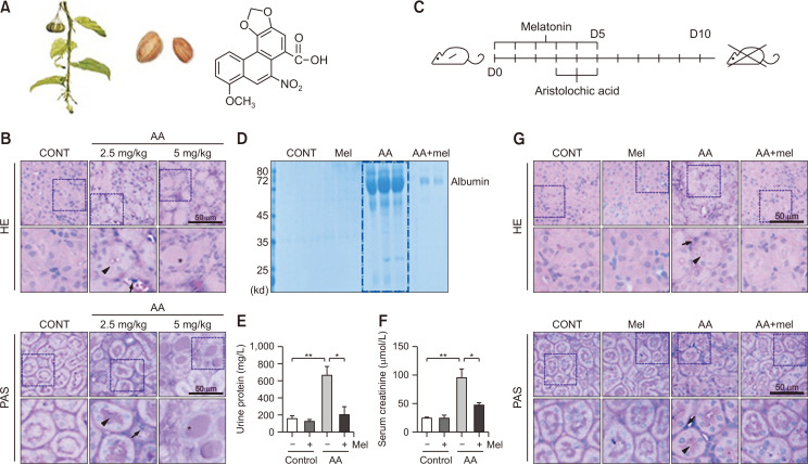
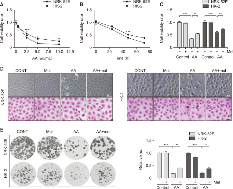
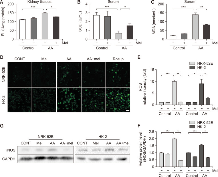
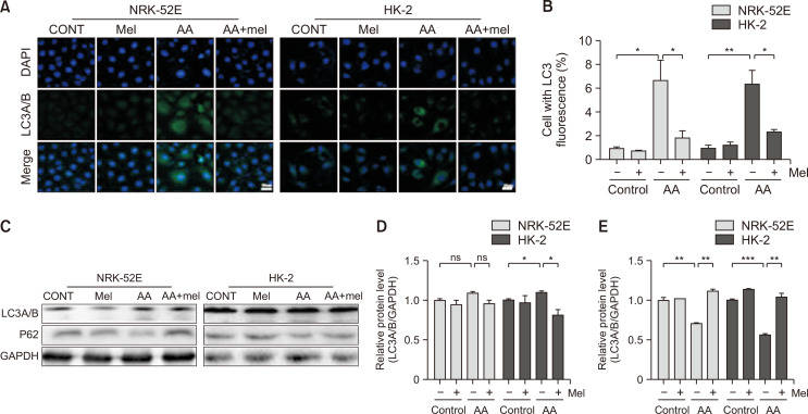
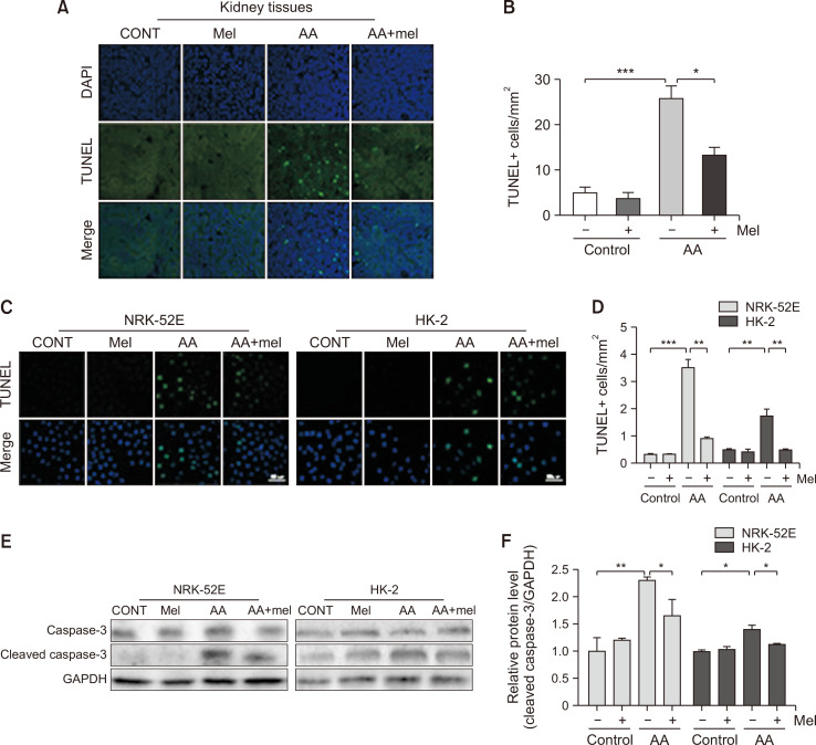
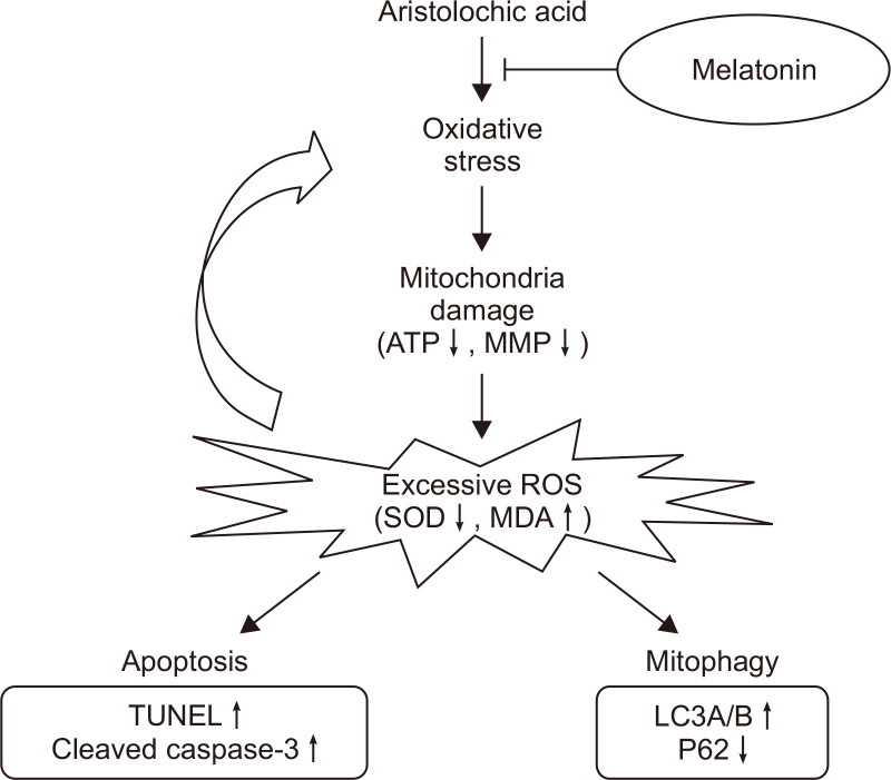
Aristolochic acid (AA), extracted from Aristolochiaceae plants, plays an essential role in traditional herbal medicines and is used for different diseases. However, AA has been found to be nephrotoxic and is known to cause aristolochic acid nephropathy (AAN). AA-induced acute kidney injury (AKI) is a syndrome in AAN with a high morbidity that manifests mitochondrial damage as a key part of its pathological progression. Melatonin primarily serves as a mitochondria-targeted antioxidant. However, its mitochondrial protective role in AA-induced AKI is barely reported. In this study, mice were administrated 2.5 mg/kg AA to induce AKI. Melatonin reduced the increase in Upro and Scr and attenuated the necrosis and atrophy of renal proximal tubules in mice exposed to AA. Melatonin suppressed ROS generation, MDA levels and iNOS expression and increased SOD activities in vivo and in vitro. Intriguingly, the in vivo study revealed that melatonin decreased mitochondrial fragmentation in renal proximal tubular cells and increased ATP levels in kidney tissues in response to AA. In vitro, melatonin restored the mitochondrial membrane potential (MMP) in NRK-52E and HK-2 cells and led to an elevation in ATP levels. Confocal immunofluorescence data showed that puncta containing Mito-tracker and GFP-LC3A/B were reduced, thereby impeding the mitophagy of tubular epithelial cells. Furthermore, melatonin decreased LC3A/B-II expression and increased p62 expression. The apoptosis of tubular epithelial cells induced by AA was decreased. Therefore, our findings revealed that melatonin could prevent AA-induced AKI by attenuating mitochondrial damage, which may provide a potential therapeutic method for renal AA toxicity.
Keywords: Acute kidney injury; Aristolochic acid; Melatonin; Mitochondrial damage; Mitophagy; Oxidative stress.
Conflict of interest statement
All authors declare that they have no conflict of interest.
Figures

Similar articles
-
Mitochondrial dysfunction is involved in aristolochic acid I-induced apoptosis in renal proximal tubular epithelial cells.Hum Exp Toxicol. 2020 May;39(5):673-682. doi: 10.1177/0960327119897099. Epub 2019 Dec 30. Hum Exp Toxicol. 2020. PMID: 31884831
-
Protective Effects of Melatonin Against Aristolochic Acid-Induced Nephropathy in Mice.Biomolecules. 2019 Dec 19;10(1):11. doi: 10.3390/biom10010011. Biomolecules. 2019. PMID: 31861726 Free PMC article.
-
The protective role of Nrf2 against aristolochic acid-induced renal tubular epithelial cell injury.Toxicol Mech Methods. 2020 Oct;30(8):580-589. doi: 10.1080/15376516.2020.1795765. Epub 2020 Aug 4. Toxicol Mech Methods. 2020. PMID: 32660364
-
Experimental Aristolochic Acid Nephropathy: A Relevant Model to Study AKI-to-CKD Transition.Front Med (Lausanne). 2022 May 4;9:822870. doi: 10.3389/fmed.2022.822870. eCollection 2022. Front Med (Lausanne). 2022. PMID: 35602498 Free PMC article. Review.
-
Environmental toxin-induced acute kidney injury.Clin Kidney J. 2017 Dec;10(6):747-758. doi: 10.1093/ckj/sfx062. Epub 2017 Jul 28. Clin Kidney J. 2017. PMID: 29225803 Free PMC article. Review.
Cited by
-
Identification and analysis of differently expressed transcription factors in aristolochic acid nephropathy.Environ Health Prev Med. 2024;29:30. doi: 10.1265/ehpm.23-00245. Environ Health Prev Med. 2024. PMID: 38777778 Free PMC article.
-
Protective effects of melatonin against physical injuries to testicular tissue: A systematic review and meta-analysis of animal models.Front Endocrinol (Lausanne). 2023 Jan 31;14:1123999. doi: 10.3389/fendo.2023.1123999. eCollection 2023. Front Endocrinol (Lausanne). 2023. PMID: 36798664 Free PMC article.
-
Regulation of renal ischemia-reperfusion injury and tubular epithelial cell ferroptosis by pparγ m6a methylation: mechanisms and therapeutic implications.Biol Direct. 2024 Oct 23;19(1):99. doi: 10.1186/s13062-024-00515-9. Biol Direct. 2024. PMID: 39444036 Free PMC article.
-
Aristolochia clematitis L. Ethanolic Extracts: In Vitro Evaluation of Antioxidant Activity and Cytotoxicity on Caco-2 Cell Line.Plants (Basel). 2024 Oct 25;13(21):2987. doi: 10.3390/plants13212987. Plants (Basel). 2024. PMID: 39519906 Free PMC article.
References
LinkOut - more resources
Full Text Sources
