. 2022 Nov:119:103394.
doi: 10.1016/j.dnarep.2022.103394. Epub 2022 Sep 6.

O-GlcNAc transferase is important for homology-directed repair

Affiliations

O-GlcNAc transferase is important for homology-directed repair

Xiaoli Ping et al. DNA Repair (Amst). 2022 Nov.

Abstract

O-Linked β-N-acetylglucosamine glycosylation (O-GlcNAcylation) to serine or threonine residues is a reversible and dynamic post-translational modification. O-GlcNAc transferase (OGT) is the only enzyme for O-GlcNAcylation, and is a potential cancer therapeutic target in combination with clastogenic (i.e., chromosomal breaking) therapeutics. Thus, we sought to examine the influence of O-GlcNAcylation on chromosomal break repair. Using a set of DNA double strand break (DSB) reporter assays, we found that the depletion of OGT, and its inhibition with a small molecule each caused a reduction in repair pathways that involve use of homology: RAD51-dependent homology-directed repair (HDR), and single strand annealing. In contrast, such OGT disruption did not obviously affect chromosomal break end joining, and furthermore caused an increase in homology-directed gene targeting. Such disruption in OGT also caused a reduction in clonogenic survival, as well as modifications to cell cycle profiles, particularly an increase in G1-phase cells. We also examined intermediate steps of HDR, finding no obvious effects on an assay for DSB end resection, nor for RAD51 recruitment into ionizing radiation induced foci (IRIF) in proliferating cells. However, we also found that the influence of OGT on HDR and homology-directed gene targeting were dependent on RAD52, and that OGT is important for RAD52 IRIF in proliferating cells. Thus, we suggest that OGT is important for regulation of HDR that is partially linked to RAD52 function.

Keywords: Double-strand break; Homology-directed repair; O-GlcNAc; OGT; RAD52.


Conflict of interest statement

Conflict of interest The authors declare that there are no conflicts of interest.

Figures
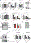
Figure 1.
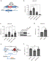
Figure 1.. OGT enzymatic activity promotes homology-directed repair and single-strand annealing.
A) Influence of OGT/OGA on distinct DSB repair pathways in U2OS human osteosarcoma cells, which are used for the entire study. Shown are the GFP+ frequencies for three DSB reporters: DR-GFP (HDR), EJ5-GFP (Distal-EJ), and SA-GFP (SSA). Cells were treated with siRNAs (pool of four) targeting OGT (siOGT-Pool), OGA (siOGA-Pool) and BRCA2 (siBRCA2-Pool). GFP+ frequencies were normalized to the parallel non-targeting siRNA (siCTRL) samples. n = 6. Shown on the right are the immunoblot signals confirming depletion of OGT and OGA. B) Influence of individual siRNA targeting OGT on distinct DSB repair pathways. GFP+ frequencies are shown as in (A), and include an siRNA targeting the end resection factor CtIP (siCtIP), as a control. n = 6. C) Depletion of OGT reduces O-GlcNAc levels. Shown are the immunoblot and dot blot signals of OGT and O-GlcNAc for cells treated with siRNAs targeting OGT. D) Influence of OSMI-4 on distinct DSB repair events. Shown are GFP+ frequencies for cells treated with OSMI-4 or DMSO. GFP+ frequencies are normalized to parallel DMSO treated samples. n = 6. Shown are dot blot signals for O-GlcNAc. E) OGT-WT expression, but not the OGT-K842M mutant, promotes HDR and SSA as well as O-GlcNAc levels in siOGT-depleted cells. Cells were treated as in (A), but expression vectors for OGT-WT and OGT-K842M were transfected together with the I-SceI expression vector. Shown are immunoblotting signals for OGT, and dot blot signals for O-GlcNAc. Shown are the GFP+ frequencies, normalized as in (A). n = 6. Error bars indicate S.D. *P≤0.05, **P≤0.01, ***P≤0.001, ****P≤0.0001, n.s.=not significant, using unpaired t-test. All experiments performed with U2OS cells.
Figure 2.
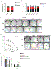
Figure 2.. OGT inhibits homology-directed gene targeting.
A) Schematic diagram of LMNA-HR assay for homology-directed gene targeting. The repair of a Cas9/sgRNA DSB in LMNA by gene targeting using the plasmid template causes integration of mRuby2 into the LMNA gene locus. B) OGT depletion causes an increase in homology-directed gene targeting. Shown are frequencies of mRuby+ cells for siRNA treated cells for the reporter in (A), normalized to transfection efficiency. n = 6. C) OGT-WT expression, but not the OGT-K842M mutant, suppresses homology-directed gene targeting in siOGT-treated cells. Cells were treated as in (B), but including OGT-WT/OGT-K842M expression vectors in the transfection with Cas9/sgRNA and the LMNA plasmid template. Shown are the mRuby+ frequencies, normalized to transfection efficiency. Also shown are immunoblot signals confirming expression of OGT. D) OSMI-4 treatment causes an increase in homology-directed gene targeting. Shown are the relative mRuby+ frequencies for the reporter in (A) for OSMI-4 treated cells normalized to transfection efficiency. n = 6. E) Shown is the schematic diagram of GAPDH-CD4 rearrangement assay for examining deletion rearrangement using endogenous genes. F) Shown are CD4+ frequencies in siRNA treated cells, normalized to siCTRL treated cells. n = 3. Error bars indicate S.D. *P≤0.05, **P≤0.01, ***P≤0.001, ****P≤0.0001, n.s.=not significant, using unpaired t-test. All experiments performed with U2OS cells.
Figure 3.
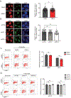
Figure 3.. OGT disruption causes an increase in G1 phase cells and reduced clonogenic survival.
A) OSMI-4 treatment causes an increase in G1 phase cells, and a decrease in G2/M phase cells. Cell cycle profiles were determined using BrdU labeling and DNA counterstain (propidium iodide). Shown is the frequency of cells in G1, S and G2/M. n = 3. B) OGT depletion by four different siRNAs causes an increase in G1 phase cells, but to differing degrees. Cell cycle profiles were determined as in (A). n = 3. C) OSMI-4 treatment causes reduced clonogenic survival. Representative images of the colony formation assays from U2OS cells that were treated with OSMI-4 for 18 hr prior to 2 Gy IR treatment or mock treatment (0 Gy), followed by continuous treatment with OSMI-4. D) Shown is the percentage clonogenic survival fraction for replicates of experiments shown in (C). Statistical comparisons are with the DMSO treated samples. Survival percentages for 2 Gy combined with 2 and 5 μM OSMI-4 were below the limit of detection. n = 3. E) OGT depletion causes reduced clonogenic survival, and expression of OGT wildtype, but not OGT-K842M promotes clonogenic survival in siOGT treated cells. Shown are the representative images of colony forming assays for cells transfected with the siRNAs and plasmids shown, and treated with 2 Gy IR or mock treated (0 Gy). F) Shown is the percentage of clonogenic survival for replicates of experiments shown in (E), n = 3. Error bars indicate S.D. *P≤0.05, **P≤0.01, ***P≤0.001, ****P≤0.0001, n.s.= not significant, using unpaired t-test. All experiments performed with U2OS cells.
Figure 4.
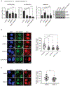
Figure 4.. OGT has no clear effect on RAD51 ionizing radiation-induced foci, nor chromosomal break end resection.
A) Ionizing radiation-induced foci (IRIF) of RAD51 is not obviously affected by OSMI-4 treatment. After OSMI-4 treatment (17 hrs), U2OS cells were labeled with EdU prior to exposure to 6 Gy IR, and 4 hours post IR treatment, cells were fixed for immunofluorescence analysis. Shown are the representative images of RAD51 and EdU staining (scale bar =10 μm). Shown are the number of RAD51 IRIF in EdU+ cells for 100 cells from two independent experiments. B) Depletion of OGT does not obviously affect RAD51 IRIF, which were analyzed as in (A), except following or siRNA treatment. Shown on are the number of RAD51 IRIF in EdU+ cells for 50 cells from two independent experiments. C) OSMI-4 treatment does not obviously affect etoposide-induced chromatin bound RPA2, which is a measure of chromosomal break end resection. After OSMI-4 treatment (or DMSO control), cells were incubated with etoposide, and mild detergent extracted prior to fixation and staining for RPA2 and DAPI. Shown are representative flow cytometry plots with the upper gate showing high RPA staining, along with frequency of RPA2+ cells from replicates of this experiment. n = 3. D) Treating cells with siOGT-1 and siOGT-2 does not obviously affect chromosomal break end resection, determined as in (C). Shown are representative flow cytometry plots and frequencies of RPA2+ cells, as in (C). n = 3, Error bars indicate S.D. n.s.=not significant. All experiments performed with U2OS cells.
Figure 5.
Figure 5.. The influence of OGT for HDR and homology-directed gene targeting is dependent on RAD52, and OGT is important for RAD52 IRIF.
A) Influence of single and combined depletion of OGT and RAD52 on HDR and SSA. Shown on the left are GFP+/ mRuby+ frequencies in cells treated with siCTRL, siOGT (siOGT-Pool), siRAD52 (siRAD52-Pool), or both siOGT and siRAD52 combined. GFP+/ mRuby+ frequencies were normalized to the parallel siCTRL treated samples. n = 6. Shown are immunoblot signals confirming depletion of OGT and RAD52. B) RAD52 IRIF are reduced in OGT depleted cells. Following siRNA treatment, U2OS cells stably expressing GFP-RAD52 were labeled with EdU for 1 hour and then was exposed to 6 Gy IR and allowed to recover for 4 hours prior to fixation for immunofluorescence analysis. Shown are representative images of RAD52 and EdU staining, and numbers of RAD52 IRIF in EdU+ cells (counted using Image J) for 50 cells from 2 independent experiments C) RAD52 IRIF are reduced in OSMI-4 treated cells. RAD52 IRIF in EdU+ cells were analyzed as in (B), following 17 hr treatment of OSMI-4, or DMSO control. Shown are representative images and numbers of RAD52 IRIF in EdU+ cells for 50 cells from 2 independent experiments. Error bars indicate S.D. *P≤0.05, **P≤0.01, ***P≤0.001, ****P≤0.0001, n.s.=not significant, using unpaired t-test. (scale bar =10 μm). All experiments performed with U2OS cells.

Similar articles

Cited by

References

    1. Dong DLY, Hart GW, Purification and Characterization of an O-Glcnac Selective N-Acetyl-Beta-D-Glucosaminidase from Rat Spleen Cytosol, J Biol Chem, 269 (1994) 19321–19330. - PubMed
    1. Hanover JA, Yu S, Lubas WB, Shin SH, Ragano-Caracciola M, Kochran J, Love DC, Mitochondrial and nucleocytoplasmic isoforms of O-linked GlcNAc transferase encoded by a single mammalian gene, Arch Biochem Biophys, 409 (2003) 287–297. - PubMed
    1. Haltiwanger RS, Holt GD, Hart GW, Enzymatic Addition of O-Glcnac to Nuclear and Cytoplasmic Proteins - Identification of a Uridine Diphospho-N-Acetylglucosamine-Peptide Beta-N-Acetylglucosaminyltransferase, J Biol Chem, 265 (1990) 2563–2568. - PubMed
    1. Hart GW, Housley MP, Slawson C, Cycling of O-linked beta-N-acetylglucosamine on nucleocytoplasmic proteins, Nature, 446 (2007) 1017–1022. - PubMed
    1. Zachara NE, Molina H, Wong KY, Pandey A, Hart GW, The dynamic stress-induced “O-GlcNAc-ome” highlights functions for O-GlcNAc in regulating DNA damage/repair and other cellular pathways, Amino Acids, 40 (2011) 793–808. - PMC - PubMed

Publication types