. 2022 Sep 20;119(38):e2205691119.
doi: 10.1073/pnas.2205691119. Epub 2022 Sep 12.

Multistate structures of the MLL1-WRAD complex bound to H2B-ubiquitinated nucleosome

Affiliations

Multistate structures of the MLL1-WRAD complex bound to H2B-ubiquitinated nucleosome

Sanim Rahman et al. Proc Natl Acad Sci U S A. .

Abstract

The human Mixed Lineage Leukemia-1 (MLL1) complex methylates histone H3K4 to promote transcription and is stimulated by monoubiquitination of histone H2B. Recent structures of the MLL1-WRAD core complex, which comprises the MLL1 methyltransferase, WDR5, RbBp5, Ash2L, and DPY-30, have revealed variability in the docking of MLL1-WRAD on nucleosomes. In addition, portions of the Ash2L structure and the position of DPY30 remain ambiguous. We used an integrated approach combining cryoelectron microscopy (cryo-EM) and mass spectrometry cross-linking to determine a structure of the MLL1-WRAD complex bound to ubiquitinated nucleosomes. The resulting model contains the Ash2L intrinsically disordered region (IDR), SPRY insertion region, Sdc1-DPY30 interacting region (SDI-motif), and the DPY30 dimer. We also resolved three additional states of MLL1-WRAD lacking one or more subunits, which may reflect different steps in the assembly of MLL1-WRAD. The docking of subunits in all four states differs from structures of MLL1-WRAD bound to unmodified nucleosomes, suggesting that H2B-ubiquitin favors assembly of the active complex. Our results provide a more complete picture of MLL1-WRAD and the role of ubiquitin in promoting formation of the active methyltransferase complex.

Keywords: MLL1; chromatin; cryo-EM; methyltransferase; ubiquitin.


Conflict of interest statement

Competing interest statement: C.W. is a member of the ThermoFisher Scientific Advisory Board.

Figures

Fig. 1.
Fig. 1.
Map of MLL1-WRAD complex shows additional density unexplained by previous structures. (A) Bar diagram depicting the MLL1-WRAD complex. Circled (not visible in map and PDB) and boxed areas represent structured domains, and white-striped boxes show newly added domains in this work. (B) Cryo-EM map of MLL1-WRAD calculated at 6-Å resolution showing fit of previously reported structure, PDB 6KIU, superimposed in density. Unaccounted-for density is shown in dashed box. (C) Zoom on unaccounted-for density indicating potential subunits that could account for it.
Fig. 2.
Fig. 2.
Cross-links of MLL1-WRAD in solution. (A) Intraprotein cross-links (purple) and interprotein cross-links (green) are indicated by lines. (B) Visualization of cross-linked lysines on the MLL1-WRAD–nucleosome complex. Satisfied (black) and violated (red) cross-links were determined using BS3′s theoretical Cα–Cα maximum crosslinking distance of 34 Å.
Fig. 3.
Fig. 3.
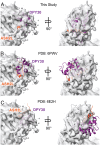
Comparison of Ash2L-SDI motif (orange) and DPY30 dimer (purple) positioning on active state map. All structures were compared by aligning the Ash2L-SPRY domain. (A) Structure of the Ash2L-SDI motif and DPY30 dimer presented in this study. (B) Structure of the Ash2L-SDI motif and DPY30 dimer reported by Park et al. (38) (6PWV) positioned in our active state map. (C) Crystal structure of the Ash2L-SDI motif and DPY30 dimer reported by Haddad et al. (33) (6E2H) positioned in our active state map.
Fig. 4.
Fig. 4.
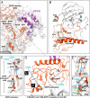
Building and fitting of the Ash2L IDR-SPRY domains to experimental density. (A) Placement of Ash2LIDR-SPRY and DPY30 model into the active state map. (B) Overview of the Ash2LIDR-SPRY model built by integrative modeling and MDFF refinement. The IDR is colored orange, and the SPRY insertion region is colored gold. Regions colored gray correspond to the SPRY domain, which was built using 6KIU as a reference model. (C) SPRY insertion stretching over 40 Å fitted to EM density. Obtained cross-links are displayed (blue, satisfied). Residues labeled in cyan refer to satisfied cross-links to the MLL1 SET domain. (D) IDR region located in the vicinity of the SPRY insertion. Intra-SPRY cross-links from K207 and K244 are displayed and demonstrate the anchoring of the IDR region. (E) Zoom on indicated region in D showing IDR loop 254 to 270 and satisfied cross-link. (F) Zoom on indicated region in D showing IDR helix 200 to 210; all cross-links are shown, lysine residues are depicted in orange, and arginine residues are depicted in green.
Fig. 5.
Fig. 5.
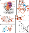
Modeling of Ash2L PHD–WH domain. (A) Model of Ash2L PhD–WH ensemble on the active state model predicted by cross-linked restrained docking. (B) Cross-links between Ash2L PhD–WH residue K67 and the Ash2L IDR and SPRY domain. (C) Additional cross-links between K99 and K122 in the Ash2L PhD–WH with K311 and K434 in the SPRY domain. (D) Single cross-link connecting Ash2L PhD–WH residue K99 to MLL1 SET domain residue K3933. (E) Zoom on indicated region in A showing the positioning the K131 and K135 in close proximity to the backbone of the nucleosomal DNA.
Fig. 6.
Fig. 6.
Four distinct states of MLL1-WRAD on the ubiquitinated nucleosome. (Top) The cartoon depictions indicate the four states discussed in the text. (Middle and Bottom) Two views of the corresponding density maps are shown beneath each cartoon, with the structure of the active state model superimposed on each map. (A) State 1, (B) state 2, (C) state 3, and (D) active state map calculated at 6 Å, shown for comparison. Densities in all panels are colored according to the cartoon diagrams.
Fig. 7.
Fig. 7.
Position of MLL1 complexes on the nucleosomes. The solvent-accessible surface is shown for each resolved subunit. Color scheme for all panels: ubiquitin, yellow; RbBP5, green; WDR5, blue; MLL1, cyan; Ash2L, orange; and DPY30, purple. (A) MLL1-WRAD complex bound to H2B-ubiquitinated nucleosome, present study (7UD5). (B) MLL1-WRAD bound to unmodified nucleosomes (6KIZ). (C) MLL-WRA bound to unmodified nucleosome (6W5M).

Similar articles

Cited by

References

    1. Jenuwein T., Allis C. D., Translating the histone code. Science 293, 1074–1080 (2001). - PubMed
    1. Ringel A. E., Cieniewicz A. M., Taverna S. D., Wolberger C., Nucleosome competition reveals processive acetylation by the SAGA HAT module. Proc. Natl. Acad. Sci. U.S.A. 112, E5461–E5470 (2015). - PMC - PubMed
    1. Bian C., et al. , Sgf29 binds histone H3K4me2/3 and is required for SAGA complex recruitment and histone H3 acetylation. EMBO J. 30, 2829–2842 (2011). - PMC - PubMed
    1. Vermeulen M., et al. , Selective anchoring of TFIID to nucleosomes by trimethylation of histone H3 lysine 4. Cell 131, 58–69 (2007). - PubMed
    1. Lauberth S. M., et al. , H3K4me3 interactions with TAF3 regulate preinitiation complex assembly and selective gene activation. Cell 152, 1021–1036 (2013). - PMC - PubMed

Publication types

MeSH terms

LinkOut - more resources