. 2022 Sep 9;8(1):90.
doi: 10.1038/s41421-022-00439-6.

Structural insights into human CCAN complex assembled onto DNA

Affiliations

Structural insights into human CCAN complex assembled onto DNA

Tian Tian et al. Cell Discov. .

Abstract

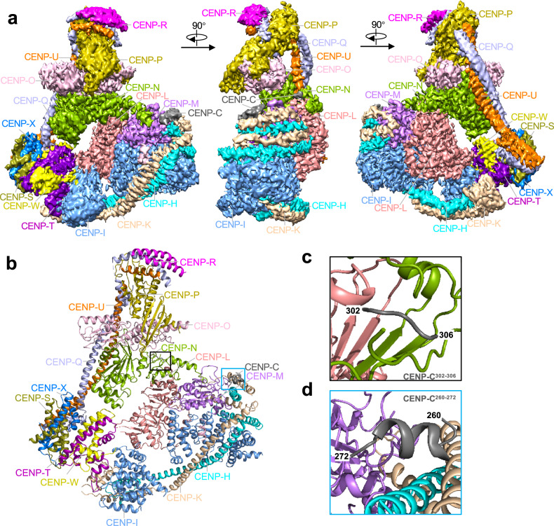
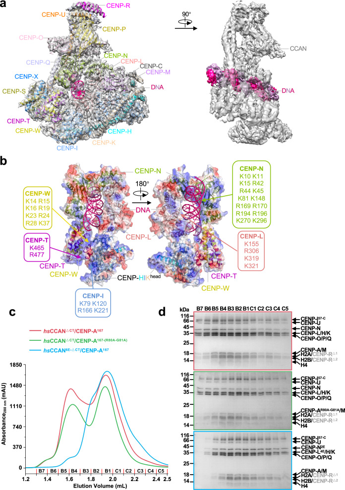
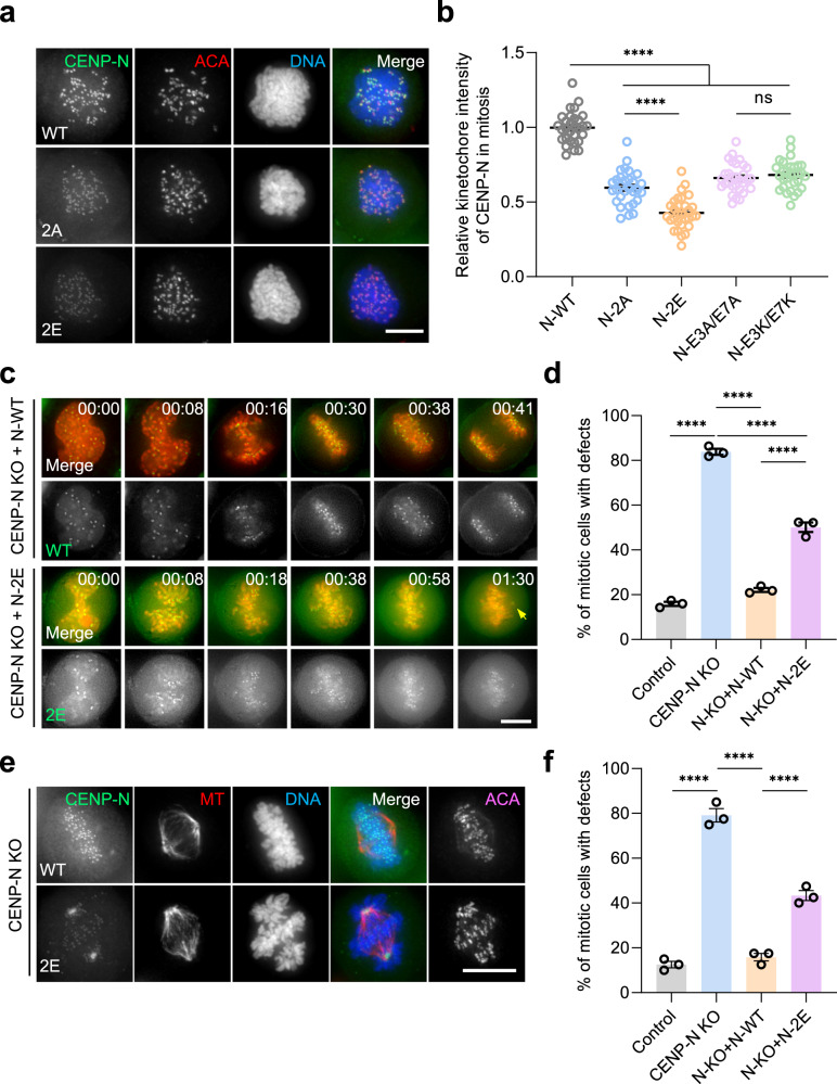
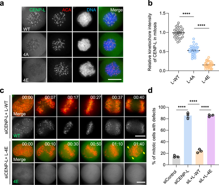
In mitosis, accurate chromosome segregation depends on kinetochores that connect centromeric chromatin to spindle microtubules. The centromeres of budding yeast, which are relatively simple, are connected to individual microtubules via a kinetochore constitutive centromere associated network (CCAN). However, the complex centromeres of human chromosomes comprise millions of DNA base pairs and attach to multiple microtubules. Here, by use of cryo-electron microscopy and functional analyses, we reveal the molecular basis of how human CCAN interacts with duplex DNA and facilitates accurate chromosome segregation. The overall structure relates to the cooperative interactions and interdependency of the constituent sub-complexes of the CCAN. The duplex DNA is topologically entrapped by human CCAN. Further, CENP-N does not bind to the RG-loop of CENP-A but to DNA in the CCAN complex. The DNA binding activity is essential for CENP-LN localization to centromere and chromosome segregation during mitosis. Thus, these analyses provide new insights into mechanisms of action underlying kinetochore assembly and function in mitosis.


Conflict of interest statement

The authors declare no competing interests.

Figures

Fig. 1
Fig. 1. Cryo-EM structure of the human CCAN complex.
a The 3.3 Å-resolution cryo-EM density map of the CCAN complex at three different views. b Cartoon representation of the CCAN structure model. All subunits are assigned into five sub-complexes, including CENP-C, CENP-LN, CENP-HIKM, CENP-OPQUR and CENP-TWSX. The black and blue box showed the fragment of CENP-C. c, d Enlarged view of the fragment of CENP-C show in b. Interface underlying CENP-C302–306 interactions with CENP-L (c, black frame). CENP-C260–272 interacts with CENP-HKM (d, blue frame). See also Supplementary Figs. S1–S4.
Fig. 2
Fig. 2. DNA binds to CCAN through the CENP-LN channel.
a The 3.7 Å resolution cryo-EM density map of CCAN–DNA complex at two different views. The density map of DNA is colored by hotpink and the others are shown in gray transparent surfaces. b Electrostatic potential surface view of CENP-LN-HIKhead-TW binding with DNA. The DNA is shown as cartoon. Note that positively charged amino acids from CENP-LN, CENP-I and CENP-TW constitute the contact sites between CCAN and DNA. c, d Comparison of elution profiles (c) of CCANΔCT-CENP-A167/CCANΔCT-CENP-A167-(R80A-G81A) and CCAN6E-ΔCT-CENP-A167 in Superose 6 5/150 GL (GE Health) and the Coomassie-blue stained 15% SDS-PAGE gel. CENP-A167 is the CENP-A nucleosome reconstituted by using a DNA fragment of 167 bp in length. d The CCANΔCT complex bound to either CENP-A nucleosome or CENP-AR80A-G81A nucleosome which reconstituted with 167 bp DNA, but the CCAN6E-ΔCT complex failed. The CCANΔCT complex includes CENP-LN, CENP-HIKM and CENP-OPQUR, but not CENP-C and CENP-TWSX; the CCAN6E-ΔCT complex includes charge mutations of positively-charged residues on the CENP-LN (K270E/K296E in CENP-N2E, K155E/R306E/K319E/K321E in CENP-L4E) in contact with DNA; two degradation products of CENP-R annotated as CENP-RΔ1 and CENP-RΔ2 in gray color. Of note, the two separated peaks seen in the elution represent wild type CCAN (red line) and CENP-ANuc–binding deficient CCAN (green line) complex with CENP-ANuc and CENP-ANuc which are indistinguishable. However, DNA binding-deficient CCAN (cyan line) failed to bind nucleosomal DNA, validating that CCAN binds to DNA via CENP-LN. See also Supplementary Figs. S5, S6.
Fig. 3
Fig. 3. DNA binding is required for CENP-N centromere localization and function in mitosis.
a Representative immunofluorescence montage of HeLa cells expressing GFP-CENP-N wild type and DNA binding-deficient mutants. Scale bar, 10 µm. Note that K270 and K296 binding to DNA determines CENP-N localization to centromere in mitosis. b Statistical analyses of centromere localization efficacy of CENP-N wild type and mutants. Data present means ± s.e.m. from three independent experiments of 30 cells for each group. Ordinary one-way ANOVA followed by Tukey’s post hoc test was used to determine statistical significance. ****p < 0.0001; ns, not significant. c Real-time imaging of HeLa cells with chromosome marked by H2B-mCherry and GFP-tagged CENP-N wild type and 2E mutant in the absence of endogenous CENP-N. Note that 2E mutant caused mitotic arrest with chromosome alignment defect. Scale bar, 10 µm. d Quantification of mitotic phenotypes in cells expressing CENP-N 2E mutant after induction of endogenous CENP-N knockout as in c. Data present means ± s.e.m. from three independent experiments (Control, n = 69; CENP-N KO, n = 68; N-KO + N-WT, n = 68; N-KO + N-2E, n = 68). Ordinary one-way ANOVA followed by Tukey’s post hoc test was used to determine statistical significance. ****p < 0.0001. e Representative immunofluorescence montage of HeLa cells expressing GFP-CENP-N wild type and 2E mutant and stained for kinetochore microtubule. Scale bar, 10 µm. Note that 2E mutant failed to localize to centromere which resulted in aberrant spindle and misaligned chromosomes. f Statistical analyses of chromosome alignment efficacy of CENP-N wild type and 2E mutant. Data present means ± s.e.m. from three independent experiments of 120 cells for each group. Ordinary one-way ANOVA followed by Tukey’s post hoc test was used to determine statistical significance. ****p < 0.0001. See also Supplementary Fig. S7.
Fig. 4
Fig. 4. CENP-L binding to DNA is essential for accurate chromosome segregation.
a Representative immunofluorescence montage of HeLa cells expressing GFP-CENP-L wild type and DNA binding-deficient mutants. Scale bar, 10 µm. Note that K155/R306/K319/K321 determine CENP-L localization to centromere in mitosis. b Statistical analyses of centromere localization efficacy of CENP-L wild type and mutants (4A, 4E). Data present means ± s.e.m. from three independent experiments of 30 cells. Ordinary one-way ANOVA followed by Tukey’s post hoc test was used to determine statistical significance. ****p < 0.0001. c Real-time imaging of HeLa cells with chromosome marked by H2B-mCherry and GFP-tagged CENP-L wild type and 4E mutant in the absence of endogenous CENP-L. Note that 4E mutant caused mitotic arrest with chromosome alignment defects. Scale bar, 10 µm. d Quantification of mitotic phenotypes in cells expressing CENP-L 4E mutant after induction of endogenous CENP-L knockout as in c. Data present means ± s.e.m. from three independent experiments of (siControl, n = 67; siCENP-L, n = 68; siL + L-WT, n = 67; siL + L-4E, n = 69). Ordinary one-way ANOVA followed by Tukey’s post hoc test was used to determine statistical significance. ****p < 0.0001. See also Supplementary Fig. S8.

Similar articles

Cited by

References

    1. Cleveland DW, Mao Y, Sullivan KF. Centromeres and kinetochores: from epigenetics to mitotic checkpoint signaling. Cell. 2003;112:407–421. doi: 10.1016/S0092-8674(03)00115-6. - DOI - PubMed
    1. McKinley KL, Cheeseman IM. The molecular basis for centromere identity and function. Nat. Rev. Mol. Cell Biol. 2016;17:16–29. doi: 10.1038/nrm.2015.5. - DOI - PMC - PubMed
    1. Rieder CL. The formation, structure, and composition of the mammalian kinetochore and kinetochore fiber. Int. Rev. Cytol. 1982;79:1–58. doi: 10.1016/S0074-7696(08)61672-1. - DOI - PubMed
    1. McIntosh JR, Grishchuk EL, West RR. Chromosome-microtubule interactions during mitosis. Annu. Rev. Cell Dev. Biol. 2002;18:193–219. doi: 10.1146/annurev.cellbio.18.032002.132412. - DOI - PubMed
    1. Foltz DR, et al. The human CENP-A centromeric nucleosome-associated complex. Nat. Cell Biol. 2006;8:458–469. doi: 10.1038/ncb1397. - DOI - PubMed