

The molecular mechanisms and therapeutic strategies of EMT in tumor progression and metastasis
- PMID: 36076302
- PMCID: PMC9461252
- DOI: 10.1186/s13045-022-01347-8
The molecular mechanisms and therapeutic strategies of EMT in tumor progression and metastasis
Abstract
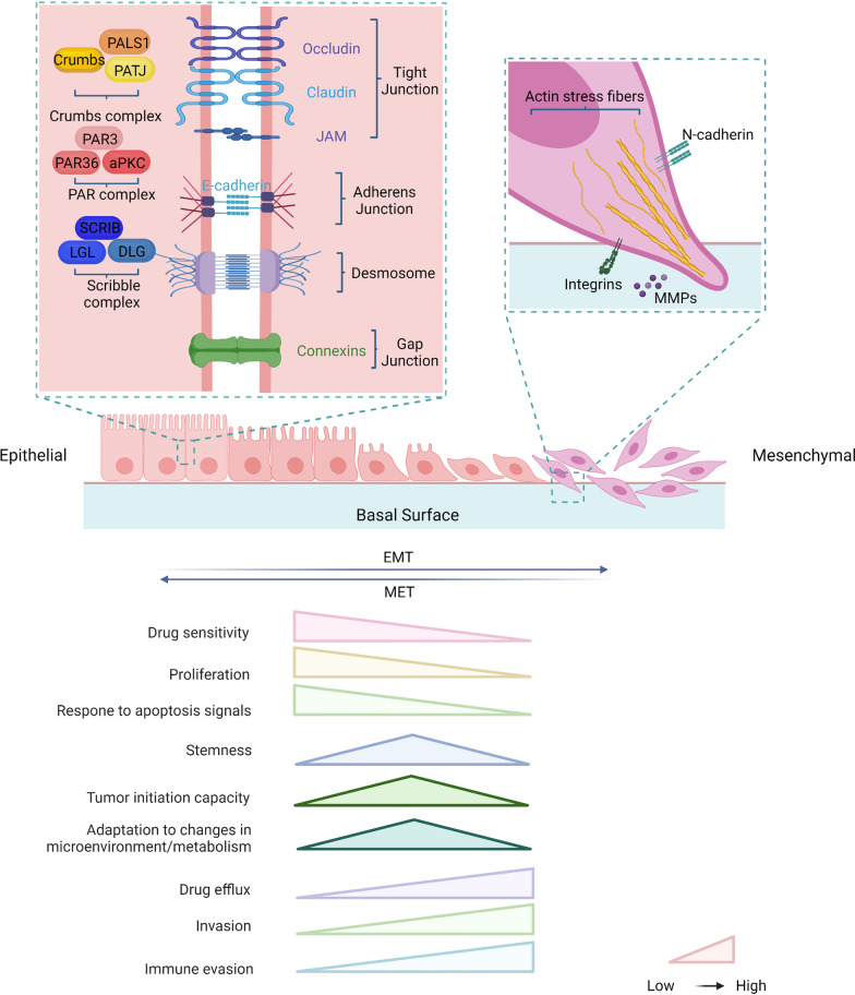
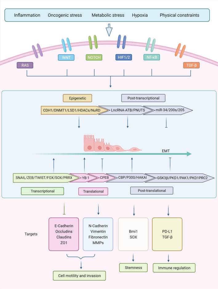
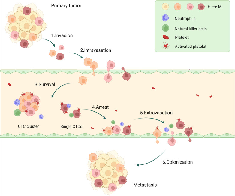
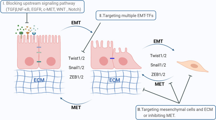
Epithelial-mesenchymal transition (EMT) is an essential process in normal embryonic development and tissue regeneration. However, aberrant reactivation of EMT is associated with malignant properties of tumor cells during cancer progression and metastasis, including promoted migration and invasiveness, increased tumor stemness, and enhanced resistance to chemotherapy and immunotherapy. EMT is tightly regulated by a complex network which is orchestrated with several intrinsic and extrinsic factors, including multiple transcription factors, post-translational control, epigenetic modifications, and noncoding RNA-mediated regulation. In this review, we described the molecular mechanisms, signaling pathways, and the stages of tumorigenesis involved in the EMT process and discussed the dynamic non-binary process of EMT and its role in tumor metastasis. Finally, we summarized the challenges of chemotherapy and immunotherapy in EMT and proposed strategies for tumor therapy targeting EMT.
Keywords: Circulating tumor cells; Epithelial–mesenchymal transition; Metastasis; Tumor stemness.
© 2022. The Author(s).
Conflict of interest statement
The authors declare no competing interests.
Figures

Similar articles
-
EMT imparts cancer stemness and plasticity: new perspectives and therapeutic potential.Front Biosci (Landmark Ed). 2021 Jan 1;26(2):238-265. doi: 10.2741/4893. Front Biosci (Landmark Ed). 2021. PMID: 33049669 Review.
-
Epithelial-Mesenchymal Plasticity in Cancer Progression and Metastasis.Dev Cell. 2019 May 6;49(3):361-374. doi: 10.1016/j.devcel.2019.04.010. Dev Cell. 2019. PMID: 31063755 Free PMC article. Review.
-
Revisiting epithelial-mesenchymal transition in cancer metastasis: the connection between epithelial plasticity and stemness.Mol Oncol. 2017 Jul;11(7):792-804. doi: 10.1002/1878-0261.12096. Epub 2017 Jun 26. Mol Oncol. 2017. PMID: 28649800 Free PMC article. Review.
-
Factors Determining Epithelial-Mesenchymal Transition in Cancer Progression.Int J Mol Sci. 2024 Aug 17;25(16):8972. doi: 10.3390/ijms25168972. Int J Mol Sci. 2024. PMID: 39201656 Free PMC article. Review.
-
Epithelial-to-mesenchymal transition in tumor progression.Med Oncol. 2017 Jul;34(7):122. doi: 10.1007/s12032-017-0980-8. Epub 2017 May 30. Med Oncol. 2017. PMID: 28560682 Review.
Cited by
-
Impact of Hypoxia and the Levels of Transcription Factor HIF-1α and JMJD1A on Epithelial-Mesenchymal Transition in Head and Neck Squamous Cell Carcinoma Cell Lines.Cancer Genomics Proteomics. 2024 Nov-Dec;21(6):591-607. doi: 10.21873/cgp.20476. Cancer Genomics Proteomics. 2024. PMID: 39467631 Free PMC article.
-
Pirfenidone inhibits TGF-β1-induced metabolic reprogramming during epithelial-mesenchymal transition in non-small cell lung cancer.J Cell Mol Med. 2024 Feb;28(3):e18059. doi: 10.1111/jcmm.18059. Epub 2023 Dec 23. J Cell Mol Med. 2024. PMID: 38140828 Free PMC article.
-
USP5 promotes tumor progression by stabilizing SLUG in bladder cancer.Oncol Lett. 2024 Sep 27;28(6):572. doi: 10.3892/ol.2024.14705. eCollection 2024 Dec. Oncol Lett. 2024. PMID: 39397799 Free PMC article.
-
AKR1C3 promotes progression and mediates therapeutic resistance by inducing epithelial-mesenchymal transition and angiogenesis in small cell lung cancer.Transl Oncol. 2024 Sep;47:102027. doi: 10.1016/j.tranon.2024.102027. Epub 2024 Jul 1. Transl Oncol. 2024. PMID: 38954974 Free PMC article.
-
Drug resistance mechanisms in cancers: Execution of pro-survival strategies.J Biomed Res. 2024 Feb 28;38(2):95-121. doi: 10.7555/JBR.37.20230248. J Biomed Res. 2024. PMID: 38413011 Free PMC article.
References
-
- Correa-Costa M, Andrade-Oliveira V, Braga TT, Castoldi A, Aguiar CF, Origassa CS, Rodas AC, Hiyane MI, Malheiros DM, Rios FJ, et al. Activation of platelet-activating factor receptor exacerbates renal inflammation and promotes fibrosis. Lab Invest. 2014;94:455–466. doi: 10.1038/labinvest.2013.155. - DOI - PubMed
Publication types
MeSH terms
Grants and funding
LinkOut - more resources
Full Text Sources
