. 2022 Sep 7;18(9):e1010385.
doi: 10.1371/journal.pgen.1010385. eCollection 2022 Sep.

Imp interacts with Lin28 to regulate adult stem cell proliferation in the Drosophila intestine

Affiliations

Imp interacts with Lin28 to regulate adult stem cell proliferation in the Drosophila intestine

Perinthottathil Sreejith et al. PLoS Genet. .

Abstract

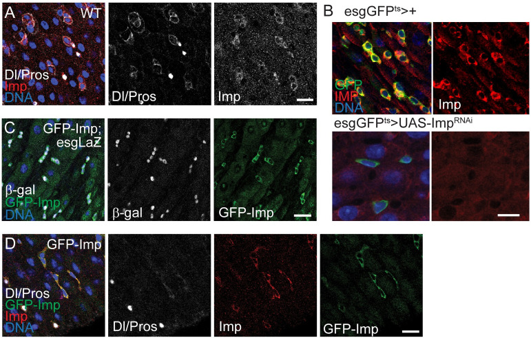
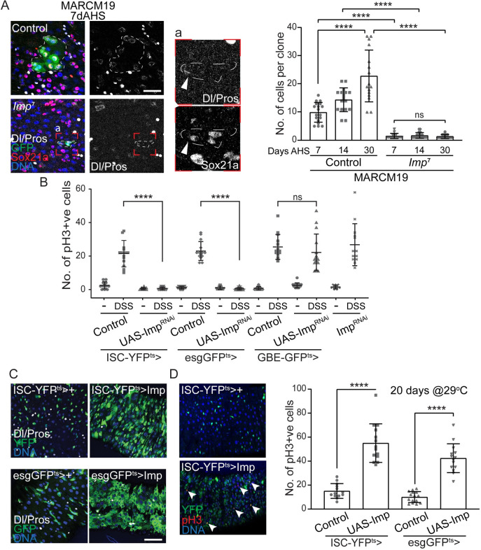
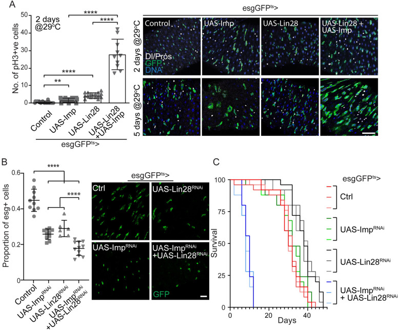
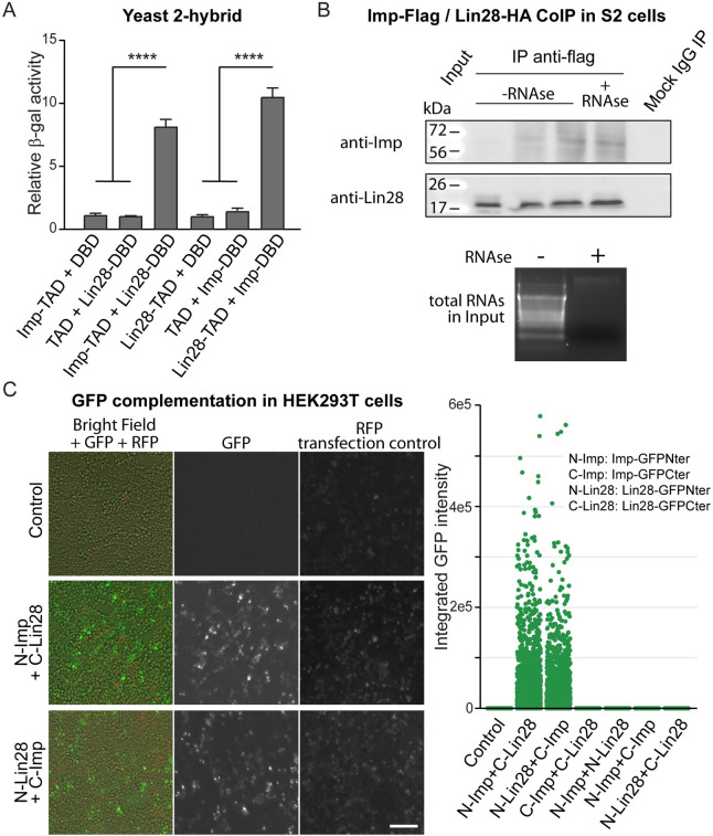
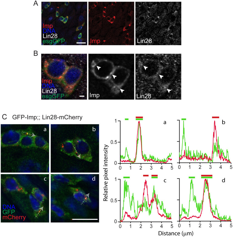
Stem cells are essential for the development and long-term maintenance of tissues and organisms. Preserving tissue homeostasis requires exquisite control of all aspects of stem cell function: cell potency, proliferation, fate decision and differentiation. RNA binding proteins (RBPs) are essential components of the regulatory network that control gene expression in stem cells to maintain self-renewal and long-term homeostasis in adult tissues. While the function of many RBPs may have been characterized in various stem cell populations, how these interact and are organized in genetic networks remains largely elusive. In this report, we show that the conserved RNA binding protein IGF2 mRNA binding protein (Imp) is expressed in intestinal stem cells (ISCs) and progenitors in the adult Drosophila midgut. We demonstrate that Imp is required cell autonomously to maintain stem cell proliferative activity under normal epithelial turnover and in response to tissue damage. Mechanistically, we show that Imp cooperates and directly interacts with Lin28, another highly conserved RBP, to regulate ISC proliferation. We found that both proteins bind to and control the InR mRNA, a critical regulator of ISC self-renewal. Altogether, our data suggests that Imp and Lin28 are part of a larger gene regulatory network controlling gene expression in ISCs and required to maintain epithelial homeostasis.


Conflict of interest statement

The authors have declared that no competing interests exist.

Figures

Fig 1
Fig 1. Imp expression in adult intestinal stem cell progenitors.
(A) Confocal images of 7-day old wild type intestinal epithelium stained with antibodies specific to Imp and Delta (Dl, ISC marker) and Prospero (Pros, enteroendocrine cell marker). (B) Confocal images of 5-day old adult guts (esg-GFPts) stained with antibody specific to Imp shows the expression of Imp in Stem cell progenitors where GFP is expressed in ISCs and EBs. Adult specific knockdown of Imp using Imp RNAi (esg-GFPts>ImpRNAi) leads to loss of Imp staining in the stem cell progenitors compared to controls (esgGFPts>+). (C) Confocal images of the intestine of 7-day old trans-heterozygote for GFP-Imp-Trap and progenitor specific Lac-Z reporter (GFP-Imp;esg-LacZ) stained with antibody specific to GFP and β-galactosidase show the expression of Imp in all LacZ-positive ISCs and EBs. (D) Confocal images of 7-day old GFP-Imp gut co-stained for delta, Prospero, Imp and GFP antibody confirms the expression of Imp in ISCs and EBs. In all panels, scale bar: 10μm.
Fig 2
Fig 2. Imp is required cell autonomously in Intestinal Stem cells.
(A) MARCM clonal analysis of control and Imp homozygous null ISCs shows that Imp is essential for ISC proliferation. Clones are labeled by GFP expression (green), Sox21a identifies ISCs and EBs, Delta (Dl) specifically stains ISCs and Prospero (Pros) highlights enteroendocrine cells. Insert ‘a’ presents a higher magnification image of an Imp mutant clone, illustrating the presence Dl-positive and Sox21a-positive cells in these very small GFP+ cell clusters. Box plot represents the distribution of number of cells per clone in an age-dependent manner. Imp mutant clones fail to grow, even after 30 days of clonal induction (days after heat shock, AHS). Student’s t-test **** p-value = < 0.0001; ns = 0.426. (B) Imp is required specifically in ISCs for cell proliferation. Cell specific knockdown of Imp in ISCs, but not in EBs, abolishes DSS- induced cell division. Proliferation is measured by the number of phospho-Histone H3 (pH3) positive cells per gut 48-hour exposure to DSS, in control animals or when Imp is knocked-down in ISCs (ISC-YFPts), ISCs+EBs (esgGFPts) or EBs (GBE-GFPts). (C, D) Over-expression of Imp in ISCs (ISC-YFPts) or ISCs+EBs (esgGFPts) is sufficient to promote cell proliferation, as shown by the expansion of the esg-positive and Dl-positive cells (C) and the increased number in pH3-positive mitotic cells (arrowheads, D). Delta (Dl) specifically stains for ISCs and Prospero (Pros) stains for EEs. DNA is stained by Hoechst. In B and D, each data point represents the number of pH3-positive cells in a gut; n>10 guts per genetic and treatment conditions. Student’s t-test **** p-value = < 0.0001; * p = <0.05; ns p = 0.3562. In panels A and C, scale bar: 10μm.
Fig 3
Fig 3. Imp and Lin28 cooperate to regulate ISC proliferation.
(A) Co-expression of Lin28 and IMP leads to synergistically increased ISC proliferation. Box plot representing the quantification of the number of pH3 positive cells per gut that shows that ISCs/EBs specific co-expression of Imp and Lin28 leads to high number of mitotic cells 2 days after transgene induction. Representative images are shown to illustrate the expansion of esg-positive cells 5 days post induction of both transgenes. GFP is expressed in ISCs and EBs, Delta stains for ISCs and Prospero (Pros) stains for EEs. DNA is stained by Hoechst. Student’s t-test **** p-value = <0.0001; ** p = 0.0036. (B) Double knockdown of Imp and Lin28 results in a long-term loss of progenitors in adult guts, 7 days after transgene activation. Plot presenting the comparison of the percentage of esg-positive in the posterior midgut (measured by the ratio of GFP positive cells vs the total number of cells, counted by DNA-positive nuclei) showing a reduced number of progenitors when either Imp or lin28 are knocked down, which is exacerbated when both Imp and Lin28 are knocked down. Representative confocal images are shown to illustrate the changes in the number of GFP-positive cells. (C) Combined loss of Imp and Lin28 reduces adult female lifespan. Adult specific knockdown of Imp and Lin28 in esg-positive cells leads to accelerated death when flies are reared at 29°C. Kaplan-Meier survival curves of three populations of 25 flies are shown for each genetic condition. In A and B, scale bar: 10μm.
Fig 4
Fig 4. Imp regulates InR mRNA expression in the intestinal epithelium.
(A) Confocal images of MARCM clones illustrating the growth of Imp7 homozygous mutant clones expressing wild-type and constitutive active forms of the insulin receptor (InR). GFP expression labels ISC clones, Sox21a stains for the progenitors and DNA is stained with Hoechst. The number of cells per clones in the different genetic backgrounds, 7 and 14 days after heat shock induction (AHS), are shown, demonstrating that activating InR signaling is sufficient to restore ISC proliferation in Imp mutants. Student’s t-test **** p-value = <0.0001 Scale bar: 10μm. (B) Relative expression of the InR mRNA, normalized to escargot, in the 2-day old gut from control animals and when Imp and/or Lin28 are expressed in esg-positive cells. Imp+Lin28 co-expression is sufficient to drive the expression of InR, suggesting that both proteins regulate InR synergistically. Student’s t-test **** p-value = <0.0001. (C) qPCR quantification of the InR mRNA, normalized to rp49, in 7-day old gut RNA extracts shows reduced InR transcript levels in Imp heterozygotes and Lin28 homozygous mutants. Student’s t-test *** p-value = 0.0005, ** p = 0.0036. (D) Representative confocal image showing the decreased levels of InR protein detected by immunostaining (green) in intestinal progenitors of Imp and Lin28 mutants, compared to wild-type animals. Dl-positive ISCs are indicated by arrowheads. DNA is stained with Hoechst. (E) Imp and Lin28 bind to InR transcripts in adult guts. 7-day old guts were subjected to RNA-immunoprecipitation using either GFP-Imp or Lin28-venus, followed by qPCR. Significant levels of InR mRNA, normalized to input materials, are pulled down using both proteins, compared to controls. Student’s t-test **** p-value = <0.0001.
Fig 5
Fig 5. Drosophila Imp and Lin28 physically interact.
(A) Imp and Lin28 can form a stable complex in yeast cells, as shown by 2-hybrid assay. Quantification of the β-galactosidase activity when Imp and Lin28 protein fusion with the Gal4 Transcriptional Activation Domain (TAD) or the LexA DNA Binding Domain (DBD). Significant reporter activity is detected only when the Imp-TAD+Lin28-DBD or Imp-DBD+Lin28-TAD protein combinations are expressed. Student’s t-test **** p-value = <0.0001. (B) Co-immunoprecipitation of HA-tagged Lin28 and FLAG-tagged Imp proteins expressed in cultured Drosophila S2 cells. Anti-Flag precipitation pulls-down both protein fusions. RNase treatment of the lysate does not affect the stability of the complex. Total RNAs stained with ethidium bromide demonstrate the efficacy of the RNase treatment. (C) Fluorescence images of GFP complementation assay in HEK293T cells show that only when the appropriate combinations of Lin28 and Imp protein fusions (GFP-ImpNter+Lin28-GFPCter or GFP-ImpCter+Lin28-GFPNter) result in significant GFP fluorescence. Quantification of the integrated GFP intensity in different combinations of Imp and Lin28 proteins is shown over hundreds of cells per condition. RFP expression serve as a transfection control and for normalization. Scale bar: 10μm.
Fig 6
Fig 6. Imp and Lin28 partially co-localize in vivo.
(A) Representative confocal image showing the co-localization of Imp and Lin28 endogenous proteins detected using specific antibodies in wildtype gut. In these 7-day old esgGFPts>GFP kept at 29°C for 5 days, GFP expression marks esg-positive ISCs and EBs. DNA is stained with Hoechst. Imp and Lin28 proteins are expressed in similar patterns in these cells. Scale bar: 10μm. (B) High magnification images of the same conditions as in (A) show both Imp and Lin28 proteins form detectable and partially overlapping foci in the cytoplasm of esg-positive cells. Scale bar: 1μm. (C) Confocal images of the intestine of GFP-Imp and Lin28-mCherry trans-heterozygotes stained with anti-GFP and anti-mCherry antibodies. Both proteins form partially overlapping cytoplasmic foci. Four groups of esg-positive cells are shown as examples. For each, the relative fluorescence intensity profiles in both GFP and mCherry channels are reported along a 5μm traced line (highlighted by the dashed line arrows). This quantification illustrates the partial correlation between the signals.

Similar articles

Cited by

References

    1. Gebauer F, Schwarzl T, Valcarcel J, Hentze MW. RNA-binding proteins in human genetic disease. Nature reviews Genetics. 2021;22(3):185–98. doi: 10.1038/s41576-020-00302-y - DOI - PubMed
    1. Hentze MW, Castello A, Schwarzl T, Preiss T. A brave new world of RNA-binding proteins. Nature reviews. 2018;19(5):327–41. doi: 10.1038/nrm.2017.130 - DOI - PubMed
    1. Degrauwe N, Suva ML, Janiszewska M, Riggi N, Stamenkovic I. IMPs: an RNA-binding protein family that provides a link between stem cell maintenance in normal development and cancer. Genes & development. 2016;30(22):2459–74. doi: 10.1101/gad.287540.116 - DOI - PMC - PubMed
    1. Jun-Hao ET, Gupta RR, Shyh-Chang N. Lin28 and let-7 in the Metabolic Physiology of Aging. Trends Endocrinol Metab. 2016;27(3):132–41. doi: 10.1016/j.tem.2015.12.006 - DOI - PubMed
    1. Conway AE, Van Nostrand EL, Pratt GA, Aigner S, Wilbert ML, Sundararaman B, et al.. Enhanced CLIP Uncovers IMP Protein-RNA Targets in Human Pluripotent Stem Cells Important for Cell Adhesion and Survival. Cell reports. 2016;15(3):666–79. doi: 10.1016/j.celrep.2016.03.052 - DOI - PMC - PubMed

Publication types

LinkOut - more resources