Review
. 2022 Aug 19:9:912848.
doi: 10.3389/fmolb.2022.912848. eCollection 2022.

Exploring the eukaryotic Yip and REEP/Yop superfamily of membrane-shaping adapter proteins (MSAPs): A cacophony or harmony of structure and function?

Affiliations
Review

Exploring the eukaryotic Yip and REEP/Yop superfamily of membrane-shaping adapter proteins (MSAPs): A cacophony or harmony of structure and function?

Timothy Angelotti. Front Mol Biosci. .

Abstract

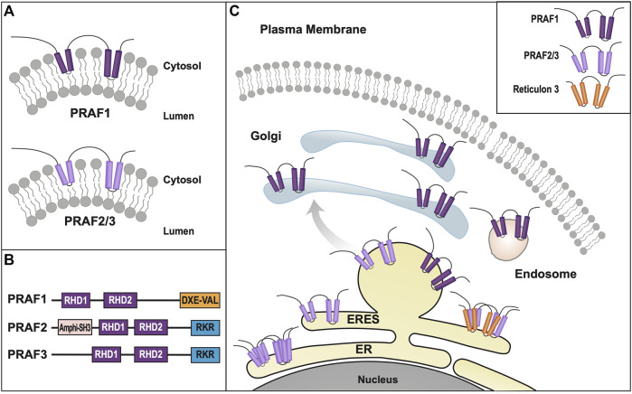
Polytopic cargo proteins are synthesized and exported along the secretory pathway from the endoplasmic reticulum (ER), through the Golgi apparatus, with eventual insertion into the plasma membrane (PM). While searching for proteins that could enhance cell surface expression of olfactory receptors, a new family of proteins termed "receptor expression-enhancing proteins" or REEPs were identified. These membrane-shaping hairpin proteins serve as adapters, interacting with intracellular transport machinery, to regulate cargo protein trafficking. However, REEPs belong to a larger family of proteins, the Yip (Ypt-interacting protein) family, conserved in yeast and higher eukaryotes. To date, eighteen mammalian Yip family members, divided into four subfamilies (Yipf, REEP, Yif, and PRAF), have been identified. Yeast research has revealed many intriguing aspects of yeast Yip function, functions that have not completely been explored with mammalian Yip family members. This review and analysis will clarify the different Yip family nomenclature that have encumbered prior comparisons between yeast, plants, and eukaryotic family members, to provide a more complete understanding of their interacting proteins, membrane topology, organelle localization, and role as regulators of cargo trafficking and localization. In addition, the biological role of membrane shaping and sensing hairpin and amphipathic helical domains of various Yip proteins and their potential cellular functions will be described. Lastly, this review will discuss the concept of Yip proteins as members of a larger superfamily of membrane-shaping adapter proteins (MSAPs), proteins that both shape membranes via membrane-sensing and hairpin insertion, and well as act as adapters for protein-protein interactions. MSAPs are defined by their localization to specific membranes, ability to alter membrane structure, interactions with other proteins via specific domains, and specific interactions/effects on cargo proteins.

Keywords: PRAF; REEP; YIPF; Yif; endoplasmic reticulum; golgi; hereditary spastic paraplegia; membrane-shaping adapter protein.


Conflict of interest statement

The author declares that the research was conducted in the absence of any commercial or financial relationships that could be construed as a potential conflict of interest.

Figures

FIGURE 1
FIGURE 1
Rab GTPase Cycle. Rab (and Ypt) GTPases cycle between inactive and active forms as they move between intraorganellar membranes (e.g. ER, Golgi) (Pfeffer and Aivazian, 2004). Such membrane cycling is dependent upon the type of guanine nucleotide bound. Left: Inactive Rab-GDP is converted to its active form by exchange of GDP for GTP, catalyzed by a guanine nucleotide exchange (GEF) factor. Inactivation of the Rab-GTP occurs by hydrolysis of GTP, which is stimulated by a GTPase-activating protein (GAP). Right: Cycling of Rab-GDP between membranes requires binding of a Rab GDP dissociation inhibitor (GDI), to prevent Rab activation as it moves through the cytoplasm. Ultimately specific membrane targeting is mediated by a membrane-bound GDI displacement factor (GDF), which releases Rab-GDP from the GDI to allow for insertion into its cognate membrane.
FIGURE 2
FIGURE 2
Yipf/Yip1p Family of Proteins. (A). When originally discovered, Yip1p was modeled as a three transmembrane domain containing protein (Yang et al., 1998). (B). Left: Alternative transmembrane topology model of Yip4p and Yip5p based on further biochemical analyses (Calero et al., 2002). Note that the membrane localization of the carboxy terminus of Yip4p/5p was determined to be intraluminal. Right: An alternative model predicted by AlphaFold (Jumper et al., 2021; Varadi et al., 2022), and yeast Yip4p/5p data (Calero et al., 2002), suggesting that their carboxy termini are closely aligned within the ER/Golgi membrane, possibly as an APH that is either aligned or buried within the membrane. (C). Yip family members have two conserved Yip1 Domains (YIPD) that may insert into the membrane as dual hairpin structures, which are necessary for Yip family homomeric and heteromeric interactions. Additionally, all members possess a carboxy terminal amphipathic helical domain (APH). (D). Intracellular localization of various Yipf family members is shown within the ER, Golgi, and ERGIC compartments, including intracellular transport vesicles, COPI and COPII. Modeled proteins are not shown to scale relative to their amino acid sequence. ER = Endoplasmic Reticulum, ERES = ER Exit Site, ERGIC = ER/Golgi Intermediate Compartment.
FIGURE 3
FIGURE 3
REEP/Yop1p Family of Proteins. (A). Domain structure of Yop1p and REEPs based on biochemical analyses (Voeltz et al., 2006; Park et al., 2010). Note that REEP1-4 and REEP5-6/Yop1p have similar structural motif but differ in their carboxy termini (Schlaitz et al., 2013). REEP/Yop1p family members have conserved Rtn homology domains (RHD) and a carboxy terminal amphipathic helical domain (APH). Additionally, REEP1-4, but not REEP5-6/Yop1p, possess a microtubule-binding domain (MTD) between RHD2 and APH (Brady et al., 2015). Additionally, REEP1-4 contain a positively charged region between RHD1 and RHD2 that also interacts with microtubules, whereas REEP5/6 possess a negatively charged region that does not interact with microtubules. (B). REEP/Yop1p family members have two conserved RHD domains that insert into the membrane as hairpin structures and a carboxy terminal APH that is either is cytoplasmic or aligned with the membrane (Park et al., 2010). (C). Intracellular localization of various REEP family members is shown within the ER, Golgi, and mitochondrial compartments, The role of REEP1 as a membrane-shaping adapter protein (MSAP) is shown with REEP1 binding to a model cargo protein (α2C adrenergic receptor) via an adapter protein (14-3-3 dimer) (Björk et al., 2013). (D). REEP1-4 possess conserved multiple potential Ser or Thr phosphorylation sites, that can bind 14-3-3 protein dimers, potential accessory proteins important for cargo trafficking (Tinti et al., 2012). Modeled proteins are not shown to scale relative to their amino acid sequence. ER = Endoplasmic Reticulum, ERES = ER Exit Site, ERGIC = ER/Golgi Intermediate Compartment.
FIGURE 4
FIGURE 4
Yif1/Yif1p Family of Proteins. (A). Transmembrane topology model of Yif1 and Yif1p based on biochemical analyses (Matern et al., 2000; Al Awabdh et al., 2012). Unlike Yipf and REEP family members, the exact Yif1 membrane topology has not been delineated but is shown as hairpin domains due to Yipf family homology. Unlike Yipf and REEP families, an APH domain has been found within the amino terminus. (B). Intracellular localization of various Yif family members is shown within the ER, Golgi, and ERGIC compartments, as well as transport machinery required for Yif1 cargo trafficking within neurons. The role of Yif1B as a regulator of cargo trafficking (and possible MSAP) is shown, interacting with a well-described cargo protein (5-HT1C receptor) (Carrel et al., 2011). Note the absence of an adapter protein. Modeled proteins are not shown to scale relative to their amino acid sequence. ER = Endoplasmic Reticulum, ERES = ER Exit Site, ERGIC = ER/Golgi Intermediate Compartment.
FIGURE 5
FIGURE 5
PRAF/Yip3p Family of Proteins. (A). Transmembrane topology model of Yip3p and PRAFs based on biochemical analyses (Abdul-Ghani et al., 2001; Lin (J). et al., 2001; Fo et al., 2006). (B). PRAF1 and PRAF2/3 have similar topologies but differ in their carboxy termini. Both have conserved YipD domains that most likely form hairpins that insert into the membrane, however they differ by the presence of either a cluster of acidic amino acid residues and a terminal Val (PRAF1) or basic amino acid residues (PRAF2/3). Potential APH domains have not been identified within this family, as has been described in other Yip families. However, an “amphiphysin Src homology 3 (SH3) group’ domain (A-SH3) has been identified in the amino terminus of PRAF2 but not PRAF3 (Lin C.-l. G. et al., 2001). (C). Intracellular localization of various PRAF family members and known binding partners is shown within the ER, Golgi, and ERGIC compartments, including intracellular transport vesicles, COPI and COPII. Modeled proteins are not shown to scale relative to their amino acid sequence. ER = Endoplasmic Reticulum, ERES = ER Exit Site, ERGIC = ER/Golgi Intermediate Compartment.
FIGURE 6
FIGURE 6
Overview of Membrane Shaping/Sensing APHs. (A). Prototypical APH (Amphipathic-Lipid-Packing-Sensor/ALPS) domain from ArfGAP1 is shown in single amino acid code and as a helical wheel diagram (yellow: bulky hydrophobic residues, purple: serines and threonines, gray: glycines and alanines, blue: positively charge residues, red: negatively charged residues) (Modified from Reference (Drin et al., 2007)). The helical wheel was generated using HELIQUEST software (Gautier et al., 2008). (B). An alternative atomistic view, demonstrating the backbone arrangement of hydrophobic (yellow) and hydrophilic (magenta) amino acid residues is also shown). Notice the facial separation of hydrophobic and hydrophilic residues which allows for the APH to either sense or associate with membrane lipids (Modified from Reference (Van Hilten et al., 2020)). (C). Similar helical wheel diagrams for APHs from other proteins that exhibit organelle membrane-specific localization are shown: Sar1 (ER membrane), endophilin 1 (plasma membrane) and ArfGAP1 (Golgi membrane) (yellow: bulky hydrophobic residues, pink: serines and threonines, white: glycines and alanines, blue: positively charge residues, red: negatively charged residues). It has been postulated that APHs are adapted to different membranes and can either induce (Sar1, endophilin) or sense (ArfGAP1) membrane curvature, depending upon the size of the hydrophobic face as well as the charge distribution within the hydrophilic face (Antonny, 2006). However, no specific amino acid sequence homology has been found to delineate these differences, suggesting that a combination of amino acid composition, spacing, and packing as well as membrane configuration may all play a part in such membrane specialization.
FIGURE 7
FIGURE 7
Caveolin and Other Membrane Shaping Adapter Proteins (MSAPs). (A). Transmembrane topology of Caveolin-1 (Cav-1). Unlike Yip family members, caveolins possess only a single hairpin domain with a carboxy terminal APH, however they meet all of the criteria to be termed MSAPs (Bauer and Pelkmans, 2006). (B). Reticulons share the most homology with REEP/Yop1p, possessing two membrane-inserted hairpin structures and a possible carboxy terminal APH (Shibata et al., 2008; Breeze et al., 2016). (C). Flotillin-1/2 are membrane-associated proteins found on the plasma membrane cytoplasmic face. They possess two hairpin structures, found within an amino terminal prohibitin-like domain (PHB) and a carboxy terminal APH (Morrow et al., 2002). (D). Protrudin, a binding partner of REEPs and atlastins, also has two hairpin domains that appear to insert into the membrane. However, instead of an APH domain, they possess a FYVE domain that discriminates between different types of phosphoinositides found in lipid rafts (Chang et al., 2013). In addition, it possesses an amino terminal ‘Rab-binding domain’ (RBD) and carboxy terminal ‘coiled-coil’ (CC) and ‘two phenylalanine’s (FF) in an acidic tract’ (FFAT) domains. (E). FAM134B, a selective ER-phagy receptor, has been recently demonstrated to possess two pairs of dual hairpin structures, with an APH domain localized between them and a carboxy terminal APH, similar to other Yipf and REEP family members (Bhaskara et al., 2019). Modeled proteins are not shown to scale relative to their amino acid sequence.

Similar articles

Cited by

References

    1. Abdul-Ghani M., Gougeon P.-Y., Prosser D. C., Da-Silva L. F., Ngsee J. K. (2001). PRA isoforms are targeted to distinct membrane compartments. J. Biol. Chem. 276 (9), 6225–6233. 10.1074/jbc.M009073200 - DOI - PubMed
    1. Abu Irqeba A., Ogilvie J. M. (2019). Novel binding partners for Prenylated Rab Acceptor 1 identified by a split-ubiquitin yeast two-hybrid screen. BMC Res. Notes 12 (1), 188. 10.1186/s13104-019-4219-y - DOI - PMC - PubMed
    1. Al Awabdh S., Miserey-Lenkei S., Bouceba T., Masson J., Kano F., Marinach-Patrice C., et al. (2012). A new vesicular scaffolding complex mediates the G-protein-coupled 5-ht1a receptor targeting to neuronal dendrites. J. Neurosci. 32 (41), 14227–14241. 10.1523/jneurosci.6329-11.2012 - DOI - PMC - PubMed
    1. AlMuhaizea M., AlMass R., AlHargan A., AlBader A., Medico Salsench E., Howaidi J., et al. (2020). Truncating mutations in YIF1B cause a progressive encephalopathy with various degrees of mixed movement disorder, microcephaly, and epilepsy. Acta Neuropathol. 139 (4), 791–794. 10.1007/s00401-020-02128-8 - DOI - PubMed
    1. Alterio J., Masson J., Diaz J., Chachlaki K., Salman H., Areias J., et al. (2015). Yif1B is involved in the anterograde traffic pathway and the Golgi architecture. Traffic 16 (9), 978–993. 10.1111/tra.12306 - DOI - PubMed

LinkOut - more resources