. 2022 Aug 17:13:942769.
doi: 10.3389/fphar.2022.942769. eCollection 2022.

Pharmacological suppression of Nedd4-2 rescues the reduction of Kv11.1 channels in pathological cardiac hypertrophy

Affiliations

Pharmacological suppression of Nedd4-2 rescues the reduction of Kv11.1 channels in pathological cardiac hypertrophy

Hua Zhang et al. Front Pharmacol. .

Abstract

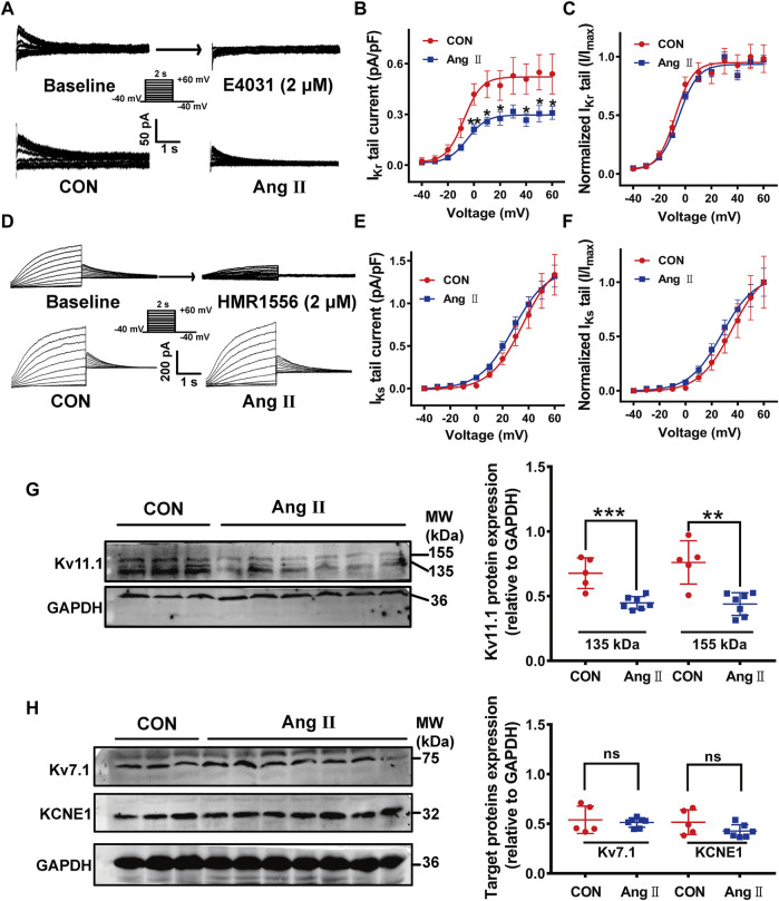
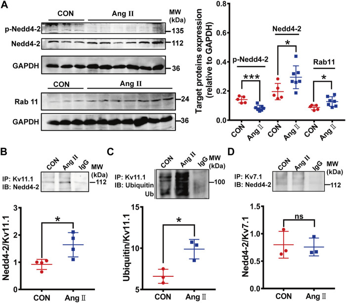
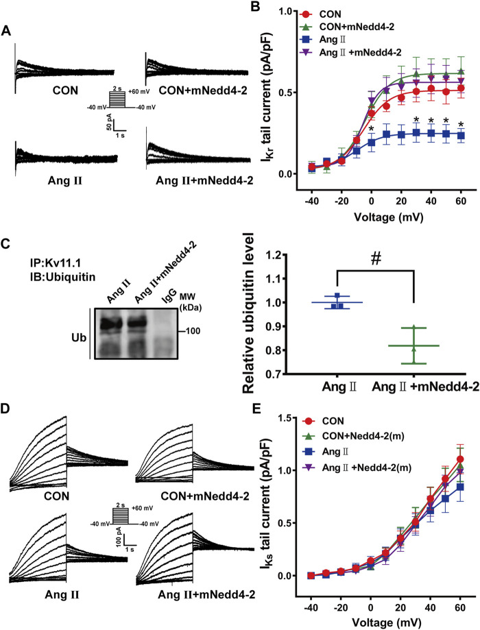
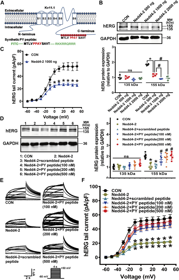
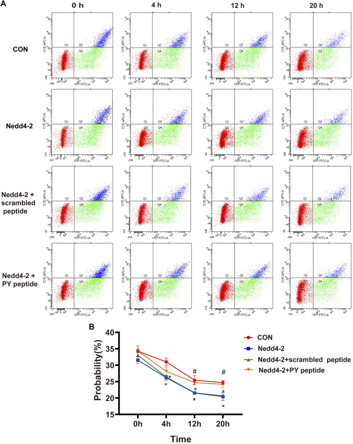
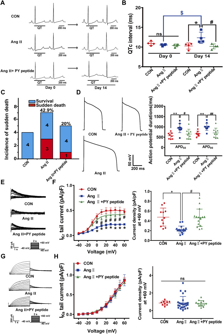
The human ether-á-go-go-related gene (hERG) encodes the pore-forming subunit (Kv11.1), conducting a rapidly delayed rectifier K+ current (I Kr). Reduction of I Kr in pathological cardiac hypertrophy (pCH) contributes to increased susceptibility to arrhythmias. However, practical approaches to prevent I Kr deficiency are lacking. Our study investigated the involvement of ubiquitin ligase Nedd4-2-dependent ubiquitination in I Kr reduction and sought an intervening approach in pCH. Angiotensin II (Ang II) induced a pCH phenotype in guinea pig, accompanied by increased incidences of sudden death and higher susceptibility to arrhythmias. Patch-clamp recordings revealed a significant I Kr reduction in pCH cardiomyocytes. Kv11.1 protein expression was decreased whereas its mRNA level did not change. In addition, Nedd4-2 protein expression was increased in pCH, accompanied by an enhanced Nedd4-2 and Kv11.1 binding detected by immunoprecipitation analysis. Cardiac-specific overexpression of inactive form of Nedd4-2 shortened the prolonged QT interval, reversed I Kr reduction, and decreased susceptibility to arrhythmias. A synthesized peptide containing the PY motif in Kv11.1 C-terminus binding to Nedd4-2 and a cell-penetrating sequence antagonized Nedd4-2-dependent degradation of the channel and increased the surface abundance and function of hERG channel in HEK cells. In addition, in vivo administration of the PY peptide shortened QT interval and action potential duration, and enhanced I Kr in pCH. We conclude that Nedd4-2-dependent ubiquitination is critically involved in I Kr deficiency in pCH. Pharmacological suppression of Nedd4-2 represents a novel approach for antiarrhythmic therapy in pCH.

Keywords: Nedd4-2; arrhythmia; pathological cardiac hypertrophy; rapid delayed rectifier K+ current; ubiquitination.


Conflict of interest statement

The authors declare that the research was conducted in the absence of any commercial or financial relationships that could be construed as a potential conflict of interest.

Figures

FIGURE 1
FIGURE 1
I Kr currents and Kv11.1 protein were reduced in the myocardium in pCH. (A) Representative tail traces of I Kr before (left) and after application of E-4031 (2 μM) (right) in upper panel. Ventricular myocyte was pulsed as shown protocol. The lower panel showing representative tail traces recorded in myocytes from control (CON) and pCH (Ang II), respectively. (B) Summary data for I Kr tail current density-voltage relationship in control and pCH (n = 10–14 cardiomyocytes from 3 to 5 hearts, *p < 0.05 versus CON). (C) Normalized current-voltage relationship for I Kr tail current. Curves were fit by Boltzmann function. (D) Representative I Ks traces recorded using the pulse protocol shown in the inset before (left) and after application of HMR1556 (2 μM) (right) in upper panel. The lower panel showing representative traces recorded in myocytes from control (CON) and pCH (Ang II), respectively. (E) Summary data for I Ks tail current density-voltage relationship in control and pCH (n = 11–15 cardiomyocytes from 3 to 5 hearts). (F) Normalized current-voltage relationship for I Ks tail current. Curves were fit by Boltzmann function. (G) Representative immunoblots bands for mature and immature Kv11.1 proteins and corresponding summary data (CON n = 5, Ang II n = 7). (H) Representative immunoblots bands for Kv7.1 and KCNE1 proteins and corresponding summary data (CON n = 5, Ang II n = 7). GADPH was used as an internal control to normalize these bands. *p < 0.05, **p < 0.01, and ***p < 0.001 versus CON. ns: not statistically significant.
FIGURE 2
FIGURE 2
Nedd4-2-mediated ubiquitination of Kv11.1 was increased in pCH. (A) Representative immunoblots showing Nedd4-2, phosphorylated Nedd4-2 (p-Nedd4-2) and Rab11 proteins and corresponding quantifications of band densities in myocardium from control (CON) (n = 5) and pCH (Ang II, n = 7). GADPH was used as an internal control to normalize the bands. (B) Representative blots immunoprecipitated (IP) by anti-Kv11.1, and western immunoblotting (IB) was performed by using anti-Nedd4-2 antibody. Quantification of band densities was shown as the ratio of Nedd4-2 to Kv11.1. (C) Representative blots immunoprecipitated by anti-Kv11.1, and western immunoblotting (IB) was performed by using anti-ubiquitin antibody. Quantification of band densities was shown as the ratio of ubiquitin to Kv11.1. (D) Proteins from control and pCH myocardium were immunoprecipitated with an anti-Kv7.1 or anti-IgG antibody, and IB was performed by using anti-Nedd4-2 antibody. IgG was used as negative controls. n = 3–4. *p < 0.05 and ***p < 0.001 versus CON. ns: not statistically significant.
FIGURE 3
FIGURE 3
Overexpression of inactive Nedd4-2 (mNedd4-2) in guinea pigs prevented Ang II-induced electrical remodeling in in vivo and ex vivo heart. (A) Schematic diagram of mNedd4-2 treatment. (B) Summary data for the incidence of sudden death. (C) Representative in vivo ECG recordings at week 6 (end of the treatment) (n = 6–10 guinea pigs each group). QT interval measurements were labeled. (D) Summary data for the quantification of QTc intervals. (E) Representative action potential traces recorded from isolated ventricular myocytes. (F) Corresponding summary data for APD50 and APD90 (n = 5-8 from 3 hearts in each group). (G) Representative ECG tracings recorded from isolated perfused hearts under burst pacing. (H) Incidence of induced ventricular tachycardia with an intensity of 120 mA. (I) The ventricular tachycardia threshold (VTT) among different groups (n = 3–4). *p < 0.05, **p < 0.01, ***p < 0.001 versus CON; # p < 0.05, ## p < 0.01, ### p < 0.001 versus Ang II; $$$ p < 0.001 versus week 0. ns: not statistically significant.
FIGURE 4
FIGURE 4
Overexpression of mNedd4-2 prevented the downregulation of Kv11.1 channels. (A) Representative I Kr tail current tracings elicited by voltage pulses shown in the inset from isolated ventricular myocytes. (B) Quantification of I Kr currents density-voltage relationship (n = 9–24 from 3 to 5 hearts in each group). (C) Representative immunoblots showed that left ventricular tissue proteins were pulled down by anti-Kv11.1 or anti-IgG antibodies and probed by anti-ubiquitin antibody (n = 3). (D) Representative I Ks currents elicited by a pulse protocol shown in the inset. (E) Summary data of I Ks currents density-voltage relationship (n = 14–30 from 3 to 5 hearts in each group). *p < 0.05 versus CON, # p < 0.05 versus Ang II.
FIGURE 5
FIGURE 5
The synthetic PY peptide prevented Nedd4-2-dependent degradation of the hERG channel expressed in hERG-HEK293 cells. (A) Diagram showing that the amino acid sequence of Nedd4-2 targeting PPAY motif is located in C-terminal region of hERG channel, and a synthetic polypeptide chain with the same sequence of 12 amino acids containing PPAY. (B) Representative immunoblots of hERG protein and quantification of the band intensities showing the effect of Nedd4-2 overexpression on the expression of hERG protein (n = 4, *p < 0.05, # p < 0.05). (C) Summary data for tail current density-voltage relationship (n = 16–26 cells in each group). (D) Representative immunoblots (left panel) and quantification of band density (right panel) (n = 5–7 samples in each group). (E) Representative hERG current tracings elicited by the voltage protocol in the inset. (F) Corresponding summary data of hERG current density- voltage relationships (n = 15–24 cells in each group). *p < 0.05 versus CON; # p < 0.05 versus Nedd4-2 alone. ns: not statistically significant.
FIGURE 6
FIGURE 6
Flow cytometry analysis of membrane hERG channels. (A) Based on the analyses of single-color controls, vertical and horizontal lines divided YFP- and BTX-CY5-positive cells, respectively. Representatives are YFP-positive cells with BTX-CY5 signal above (blue dots) or below threshold (green dots) and untransfected cells (red dots). (B) Time course of BBS-hERG-YFP delivery to the surface. The curves show probability in different times of 0, 4, 12 and 20 h treatment on BBS-hERG-YFP-HEK293 cells with BFA (n = 3). *p < 0.05 versus CON, # p < 0.05 versus Nedd4-2.
FIGURE 7
FIGURE 7
The synthetic PY peptide prevented cardiac electrical remodeling in pCH guinea pigs. (A) Representative in vivo ECG recordings. QT interval measurements were labeled. (B) Summary data for the quantification of QTc intervals (n = 4 in each group). (C) Incidence of sudden death of animals. (D) Representative raw tracings of action potentials recorded in isolated ventricular cardiacmyocytes (left panel) and corresponding summary data for APD50 and APD90 (n = 8–10 myocytes from 4 hearts in each group). (E) Representative I Kr tail current traces recorded under a pulse protocol in inset. (F) Corresponding summary data for I Kr tail current density-voltage relationship followed by I Kr tail current densities at +60 mV (n = 11–17 myocytes recorded from 4 hearts in each group). (G) Representative I Ks current traces recorded under pulse protocol shown in inset. (H) Summary data of I Ks tail currents density-voltage relationship followed by I Ks tail current density at +60 mV (n = 10–24 cardiomyocytes recorded from 4 hearts in each group). *p < 0.05, **p < 0.01 versus CON; # p < 0.05, ## p < 0.01 versus Ang Ⅱ; $ p < 0.05 versus day 0. ns: not statistically significant.

Similar articles

Cited by

References

    1. Andersen M. N., Hefting L. L., Steffensen A. B., Schmitt N., Olesen S. P., Olsen J. V., et al. (2015). Protein kinase A stimulates Kv7.1 surface expression by regulating Nedd4-2-dependent endocytic trafficking. Am. J. Physiol. Cell. Physiol. 309 (10), C693–C706. 10.1152/ajpcell.00383.2014 - DOI - PMC - PubMed
    1. Aromolaran A. S., Subramanyam P., Chang D. D., Kobertz W. R., Colecraft H. M. (2014). LQT1 mutations in KCNQ1 C-terminus assembly domain suppress IKs using different mechanisms. Cardiovasc. Res. 104 (3), 501–511. 10.1093/cvr/cvu231 - DOI - PMC - PubMed
    1. Asbun J., Villarreal F. J. (2006). The pathogenesis of myocardial fibrosis in the setting of diabetic cardiomyopathy. J. Am. Coll. Cardiol. 47 (4), 693–700. 10.1016/j.jacc.2005.09.050 - DOI - PubMed
    1. Bezzerides V. J., Zhang A., Xiao L., Simonson B., Khedkar S. A., Baba S., et al. (2017). Inhibition of serum and glucocorticoid regulated kinase-1 as novel therapy for cardiac arrhythmia disorders. Sci. Rep. 7 (1), 346. 10.1038/s41598-017-00413-3 - DOI - PMC - PubMed
    1. Cai Y., Wang Y., Xu J., Zuo X., Xu Y. (2014). Down-regulation of ether-a-go-go-related gene potassium channel protein through sustained stimulation of AT1 receptor by angiotensin II. Biochem. Biophys. Res. Commun. 452 (3), 852–857. 10.1016/j.bbrc.2014.09.014 - DOI - PubMed