

"One Shot" Sample Evaluation of 22G, 22G upgraded, 21G and 19G needle for Endobronchial Ultrasound-EBUS-TBNA
- PMID: 36046659
- PMCID: PMC9414030
- DOI: 10.7150/jca.74022
"One Shot" Sample Evaluation of 22G, 22G upgraded, 21G and 19G needle for Endobronchial Ultrasound-EBUS-TBNA
Abstract
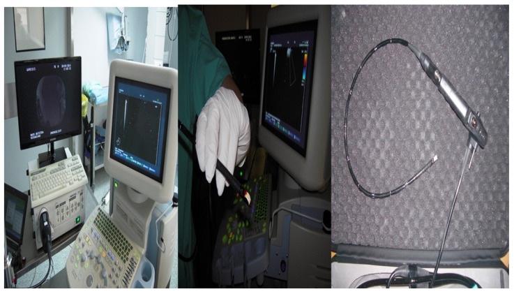
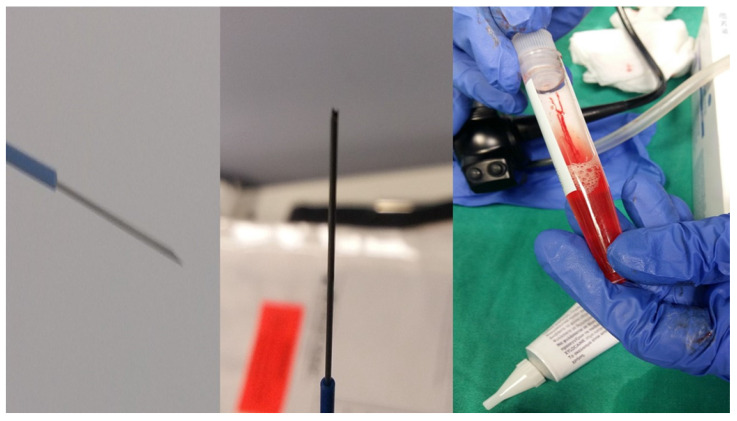
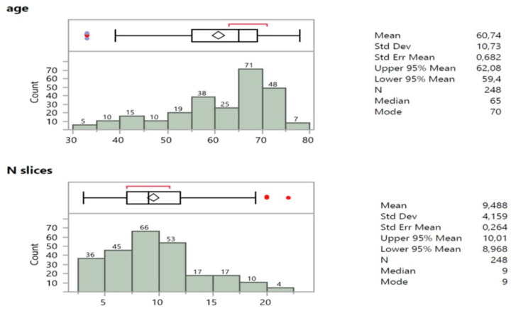
Background: There are still diagnostic issues with lung cancer and mediastinum lymphadenopathy. Endobronchial ultrasound (EBUS) is a state of the art equipment for the diagnosis of lymphadenopathy and central lesions. Objective: To investigate the sample size with one pass. Patients and Methods: 248 Stage IV patients were included in our study. All patients had a CT of the thorax with either lymphadenopathy or lyphadenopathy plus pulmonary lesions. Patients had a biopsy with endobronchial ultrasound with 22G Mediglope, 22G Mediglope Sonotip, 21G Olympus and 19G Olympus needle. We collected information regarding the cancer type, cell block, tissue, age, sex, lesion size and needle type. Results: The cancer type diagnosis was associated with the needle diameter. The number of cell-blocks were associated with the lesion size and needle diameter. Slices from the tissue and cell-blocks were again associated with the lesion size and needle diameter. Conclusion: One pass is enough for cancer diagnosis, however; careful selection has to be made among patients regarding the needle diameter. In the case of lymphoma suspicion we should use 19G needle.
Keywords: 19G needle; 21G needle; 22G needle; EBUS-TBNA; biopsy; bronchoscopy; lung cancer; lymphnodes.
© The author(s).
Conflict of interest statement
Competing Interests: The authors have declared that no competing interest exists.
Figures

Similar articles
-
Cryo-Biopsy versus 19G needle versus 22G needle with EBUS-TBNA endoscopy.J Cancer. 2022 Aug 8;13(10):3084-3090. doi: 10.7150/jca.75589. eCollection 2022. J Cancer. 2022. PMID: 36046658 Free PMC article.
-
Endobronchial Ultrasound Transbronchial Needle Aspiration With a 19-Gauge Needle vs 21- and 22-Gauge Needles for Mediastinal Lymphadenopathy.Chest. 2022 Sep;162(3):712-720. doi: 10.1016/j.chest.2022.03.041. Epub 2022 Apr 2. Chest. 2022. PMID: 35381259
-
Priority of PET-CT vs CT Thorax for EBUS-TBNA 22G vs 19G: Mesothorax Lymphadenopathy.J Cancer. 2021 Aug 5;12(19):5874-5878. doi: 10.7150/jca.59892. eCollection 2021. J Cancer. 2021. PMID: 34476000 Free PMC article.
-
Effect of Needle Size on Diagnosis of Sarcoidosis with Endobronchial Ultrasound-guided Transbronchial Needle Aspiration: Systematic Review and Meta-Analysis.Ann Am Thorac Soc. 2022 Feb;19(2):279-290. doi: 10.1513/AnnalsATS.202103-366OC. Ann Am Thorac Soc. 2022. PMID: 35103562 Review.
-
The Successful Removal of a Broken Needle as an Unusual Complication of Endobronchial Ultrasound-guided Transbronchial Needle Aspiration (EBUS-TBNA): A Case Report and Literature Review.J UOEH. 2019;41(1):35-40. doi: 10.7888/juoeh.41.35. J UOEH. 2019. PMID: 30867398 Review.
Cited by
-
Immunotherapy and Chemotherapy Versus Sleep Disturbances for NSCLC Patients.Curr Oncol. 2023 Feb 6;30(2):1999-2006. doi: 10.3390/curroncol30020155. Curr Oncol. 2023. PMID: 36826116 Free PMC article.
References
LinkOut - more resources
Full Text Sources
