. 2022 Dec;16(12):2763-2774.
doi: 10.1038/s41396-022-01310-y. Epub 2022 Aug 31.

Viral cross-class transmission results in disease of a phytopathogenic fungus

Affiliations

Viral cross-class transmission results in disease of a phytopathogenic fungus

Yue Deng et al. ISME J. 2022 Dec.

Abstract

Interspecies transmission of viruses is a well-known phenomenon in animals and plants whether via contacts or vectors. In fungi, interspecies transmission between distantly related fungi is often suspected but rarely experimentally documented and may have practical implications. A newly described double-strand RNA (dsRNA) virus found asymptomatic in the phytopathogenic fungus Leptosphaeria biglobosa of cruciferous crops was successfully transmitted to an evolutionarily distant, broad-host range pathogen Botrytis cinerea. Leptosphaeria biglobosa botybirnavirus 1 (LbBV1) was characterized in L. biglobosa strain GZJS-19. Its infection in L. biglobosa was asymptomatic, as no significant differences in radial mycelial growth and pathogenicity were observed between LbBV1-infected and LbBV1-free strains. However, cross-species transmission of LbBV1 from L. biglobosa to infection in B. cinerea resulted in the hypovirulence of the recipient B. cinerea strain t-459-V. The cross-species transmission was succeeded only by inoculation of mixed spores of L. biglobosa and B. cinerea on PDA or on stems of oilseed rape with the efficiency of 4.6% and 18.8%, respectively. To investigate viral cross-species transmission between L. biglobosa and B. cinerea in nature, RNA sequencing was carried out on L. biglobosa and B. cinerea isolates obtained from Brassica samples co-infected by these two pathogens and showed that at least two mycoviruses were detected in both fungal groups. These results indicate that cross-species transmission of mycoviruses may occur frequently in nature and result in the phenotypical changes of newly invaded phytopathogenic fungi. This study also provides new insights for using asymptomatic mycoviruses as biocontrol agent.


Conflict of interest statement

The authors declare no competing interests.

Figures
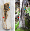
Fig. 1
Fig. 1. Field symptoms of Brassica crops co-infected by Botrytis cinerea and Leptosphaeria biglobosa.
a Co-infection of two pathogens on stem of Br. napus and b co-infection of two pathogens on petiole of Br. juncea var. tumida. Red and blue arrowheads indicate the presence of conidia of B. cinerea and pycnidia of L. biglobosa on plant tissues, respectively.
Fig. 2
Fig. 2. Genomic organization, virus particles, viral dsRNAs, and viral structure proteins of Leptosphaeria biglobosa botybirnavirus 1 (LbBV1).
a Agarose gel electrophoresis of dsRNAs extracted from the mycelium of Leptosphaeria biglobosa strain GZJS19. Marker, DNA marker D10000 (TaKaRa). b Schematic diagram of the genetic organization of LbBV1. The coding strand of dsRNA-1 is 6190 bp long and comprises one large ORF, designated ORF 1, which encodes a polyprotein with a calculated molecular mass of 202 kDa. The coding strand of dsRNA-2 is 5900 bp long and also comprises one large ORF, designated ORF 2, which encodes a polyprotein with a calculated molecular mass of 192 kDa. c Phylogenetic analysis of the RNA-dependent RNA polymerase (RdRp) region of LbBV1 and selected dsRNA viruses of Chrysoviridae, Megabirnaviridae, “Botybirnavirus” and Victoriviridae. The red dot denotes the position of LbBV1. Numbers at the nodes indicate the bootstrap values out of 1000 replicates. d Transmission electron microscopy (TEM) images of the virus particles (~37 nm in diameter) purified from L. biglobosa strain GZJS19. Arrowheads indicate the virus particles. e Agarose gel electrophoresis analysis of the dsRNAs extracted from purified virus particles of LbBV1 and the mycelia of L. biglobosa strain GZJS19. Note that the two dsRNAs of LbBV1 were not separated. Marker, DNA marker D10000 (TaKaRa). f SDS-PAGE analysis of structural proteins extracted from purified virus particles of LbBV1.
Fig. 3
Fig. 3. Biological properties of different Leptosphaeria biglobosa strains isolated from the fields.
a Colony morphology (23 °C, 8 days) of strains GZJS19, Lb731, Lb681, Lb1176, and Lb1168 on potato dextrose agar (PDA) and V8 juice agar (V8). b Radial mycelial growth rate (23 °C) on PDA and V8. c Pathogenicity assay (23 °C,7 days) of strains GZJS19, Lb731, Lb681, Lb1176, and Lb1168 on cotyledon of oilseed rape. d Lesion diameter (23 °C, 7 days) on cotyledon of rapeseed of strains GZJS19, Lb731, Lb681, Lb1176, and Lb1168. The results are expressed as arithmetic means the standard errors of the means. In each histogram, bars labeled with the same letters are not significantly different (p > 0.05) according to the least-significant-difference test.
Fig. 4
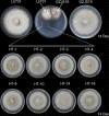
Fig. 4. Horizontal transmission of LbBV1 from Leptosphaeria biglobosa strain GZJS19 to strain Lb731 by using pairing culture technique.
The mycelium in the area of the dashed box were picked out to establish the derivative strains. The colony morphology (23 °C, 14 days) on potato dextrose agar of eight derivative strains used for biological property assays.
Fig. 5
Fig. 5. Biological properties of derivative Leptosphaeria biglobosa strains obtained from the colony of strain Lb731 after pair-culturing with strain GZJS19.
a RT-PCR detection of dsRNA-1/dsRNA-2 presence in all derivative strains by using specific primers. b Dectection of dsRNAs extracted from the mycelia all derivative strains. Marker, DNA marker D10000 (TaKaRa). c Simple sequence repeats (SSR) analysis of strains GZJS19, Lb731, and all derivative strains by using agarose gel electrophoresis. d Pathogenicity assay (23 °C, 7 days) of strains GZJS19, Lb731, and derivative strains (HT-1, HT-2, HT-3, HT-4, HT-8, HT-10, HT-14, and HT-16) on cotyledons of oilseed rape. e Radial mycelial growth rate (23 °C) of strains GZJS19, Lb731, and derivative strains on PDA. f Lesion diameter (23 °C, 7 days) of strains GZJS19, Lb731, and derivative strains on cotyledons of oilseed rape. The results are expressed as arithmetic means the standard errors of the means. In each histogram, bars labeled with the same letters are not significantly different (p > 0.05) according to the least-significant-difference test.
Fig. 6
Fig. 6. Biological effects of Leptosphaeria biglobosa botybirnavirus 1 (LbBV1) on Botrytis cinerea strain t-459.
a RT-PCR detection of dsRNA-1/dsRNA-2 presence in t-459-V with specific primers. b Agarose gel electrophoresis and northern blotting detection of dsRNAs extracted from the mycelia of L. biglobosa strains (GZJS19 and GZJS19-VF) and B. cinerea strains (t-459-V and t-459). c RT-PCR detection of the presence of dsRNA-1/dsRNA-2 in L. biglobosa and B. cinerea strains obtained from the co-culture and co-inoculation tests of L. biglobosa strain GZJS19 and B. cinerea strain t-459. The identities of these strains were confirmed by using PCR with species-specific primers, Bc = B. cinerea, Lb = L. biglobosa. Marker, DNA marker D10000 (TaKaRa). d Pathogenicity assay (20 °C, 3 days) of strains t-459 and t-459-V on detached Nicotiana benthamiana leaves. e Lesion diameter (20 °C, 3 days, lower) on detached N. benthamiana leaves of strains t-459 and t-459-V. “**” indicates a significant difference (p < 0.01) between strains t-459 and t-459-V in pathogenicity. f Colony morphology (20 °C, 20 days) of strains t-459 and t-459-V on potato dextrose agar (PDA). g Radial mycelial growth rate (20 °C, upper) of strains t-459 and t-459-V on PDA.
Fig. 7
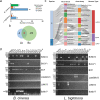
Fig. 7. The diversity of mycoviruses detected by RNA-sequencing in Leptosphaeria biglobosa and Botrytis cinerea strains isolated from co-infection samples of Brassica crops.
a The number of mycoviruses belonging to different genome types presents in each fungal population. b Venn diagram showed the number of mycoviruses present in L. biglobosa (Lb) and B. cinerea (Bc) populations. c Sankey diagram displaying the compositions of mycovirome from the populations of L. biglobosa and B. cinerea. d Detection of the presence of six mycoviruses in L. biglobosa and B. cinerea isolates by RT-PCR using the specific primers listed in Table S7. Note that two mycoviruses, Botrytis cinerea umbra-like virus 1 (BcUV1) and Botrytis cinerea mitovirus 4 (BcMV4), were detected in both two fungal populations.
Fig. 8
Fig. 8. A possible explanation of the cross species transmission between two phytopathogenic fungi, fungus A and B, sharing no similar ecological niches.
Fungus C has a wide host range and can infect both plants A and B. During the infection process, fungus C may infect plant A along with fungus A, the host-specific pathogen of plant A, at the same infection site. Thus, viruses may be able to be transmitted between A and C, no matter from A to C, or the opposite. Similarly, this case may also occur between fungus B and C, of which fungus B is the host-specific pathogen of plant B. As fungus C can be transmitted between plants A and B, it may play as a link or bridge helping the viral transmission between fungus A and B.

Similar articles

Cited by

References

    1. Casadevall A, Pirofski L-A. Benefits and costs of animal virulence for microbes. mBio. 2019;10:8–19.. - PMC - PubMed
    1. May RM, Anderson RM. Epidemiology and genetics in the coevolution of parasites and hosts. Proc R Soc B. 1983;219:281–313. - PubMed
    1. Ewald PW. Transmission modes and evolution of the parasitism-mutualism continuum. Ann NY Acad Sci. 1987;503:295–306. - PubMed
    1. Levin BR. The evolution and maintenance of virulence in microparasites. Emerg Infect Dis. 1996;2:93–101.. - PMC - PubMed
    1. Brown MJF, Loosli R, Schmid-Hempel P. Condition-dependent expression of virulence in a trypanosome infecting bumblebees. Oikos. 2000;91:421–7.

Publication types

Supplementary concepts