. 2023 Mar;17(1):89-102.
doi: 10.1007/s12079-022-00684-0. Epub 2022 Aug 30.

Chondroitin polymerizing factor predicts a poor prognosis and promotes breast cancer progression via the upstream TGF-β1/SMAD3 and JNK axis activation

Affiliations

Chondroitin polymerizing factor predicts a poor prognosis and promotes breast cancer progression via the upstream TGF-β1/SMAD3 and JNK axis activation

Qiang-Feng Pan et al. J Cell Commun Signal. 2023 Mar.

Abstract

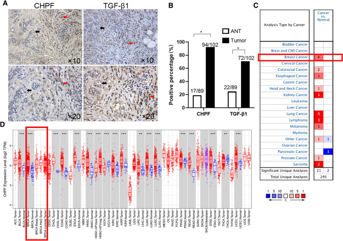
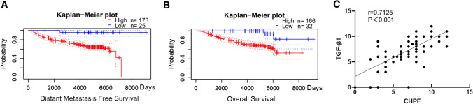
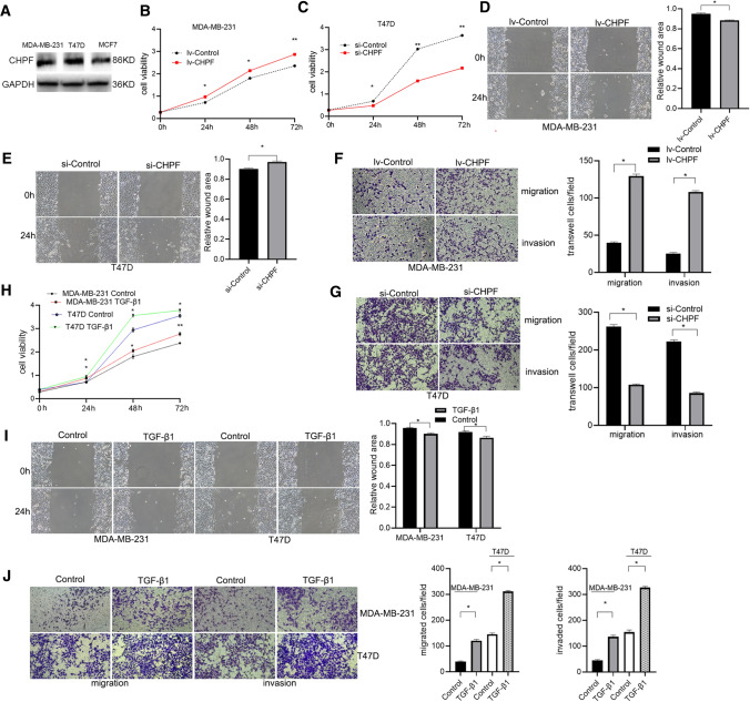
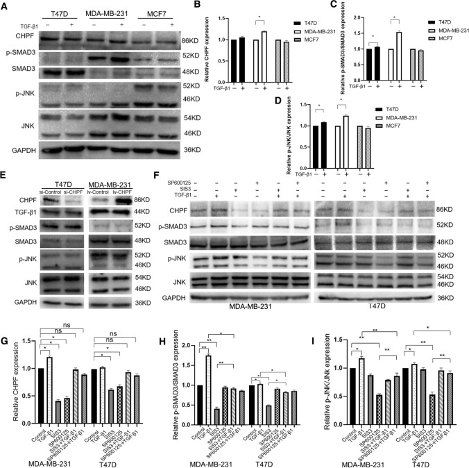
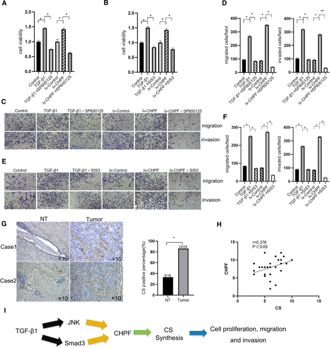
Aberrant composition of glycans in the tumor microenvironment (TME) contributes to tumor progression and metastasis. Chondroitin polymerizing factor (CHPF) is a glycosyltransferase that catalyzes the biosynthesis of chondroitin sulfate (CS). It is also correlated to transforming growth factor-β1 (TGF-β1) expression, a crucial mediator in the interaction of cancer cells with TME. In this study, we investigated the association of CHPF expression with the clinicopathological features of breast cancer (BRCA), as well the oncogenic effect and the underling mechanisms of CHPF upon BRCA cells. We found that CHPF expression is significantly increased in human BRCA tissues, and it is positively associated with TGF-β expression (r = 0.7125). The high-expression of CHPF predicts a poor prognosis and is positively correlated with tumor mass, lymph node metastasis, clinical staging and HER-2 negative-expression. The mechanistic study revealed that it promotes BRCA cell proliferation, migration and invasion through TGF-β1-induced SMAD3 and JNK activation in vitro, JNK (SP600125) or SMAD3 (SIS3) inhibitor can remove the promotion of CHPF upon cell proliferation, migration and invasion in MDA-MB-231 cells, which is derived from triple-negative breast cancer (TNBC). Collectively, our finding suggested CHPF may function as an oncogene and is highly expressed in human BRCA tissues. Pharmacological blockade of the upstream of JNK or SMAD3 signaling may provide a novel therapeutic target for refractory TNBC patients with CHPF abnormal high-expression.

Keywords: CHPF; Human breast cancer; JNK signaling; MDA-MB-231 cell line; SMAD3 signaling; TGF-β1.


Conflict of interest statement

No potential conflicts of interest were disclosed.

Figures

Fig. 1
Fig. 1
The expressions of CHPF and TGF-β1 are increased in human breast cancer tissues. a The representative IHC images of CHPF and TGF-β1 staining were showed. The black arrow indicates adjacent non-tumor tissue, the red arrow indicates BRCA tumor tissues in the images. “ × 10” and “ × 20” represent the image magnification. b The bar histogram was showed and each bar represents the positive percentage of CHPF or TGF-β1 expression (Chi-square test, “*” represents p < 0.05). “ANT” represents adjacent non-tumor tissue; “Tumor” represents BRCA tissues. c The mRNA expression of CHPF in various human tumor tissues and these corresponding normal tissues. The red square highlights the high-expression of CHPF in the BRCA tissue. Data is from Oncomine datasets. d The mRNA expression of CHPF in various human tumor tissues and normal tissues. The red square highlights the expression of CHPF in breast cancer with diverse histological subtypes. Data is from Tumor IMmune Estimation Resource (TIMER)
Fig. 2
Fig. 2
The high-expression of CHPF predicts the poor prognosis in BRCA patients and is correlated with TGF-β1 high-expression. a The high-expression of CHPF predicts the poor distant metastasis free survival in BRCA patients. Data is from PrognoScan database (COX, p < 0.05, HR 1.77 (1.13—2.76)). b The high-expression of CHPF predicts the poor overall survival in BRCA patients. Data is from PrognoScan database (COX, p < 0.05, HR 1.62 (1.02–2.56)). c CHPF and TGF-β1 expression was assessed using IHC method in BRCA tissues from our institution. The high expression of CHPF is positively correlated with TGF-β1 high-expression in BRCA tissues (Pearson linear analysis, “r” represents correlation coefficient, p < 0.001)
Fig. 3
Fig. 3
Both CHPF and TGF-β1 increase the proliferation, migration and invasion in BRCA cells Western blotting assessed the endogenous CHPF expression in a panel of T47D, MCF7 and MDA-MB-231 BRCA cell lines. B ~ G. CHPF was overexpressed (B, D and F) in MDA-MB-231 cells and knocked down (C, E and G) inT47D cells using lentivius interference techique. B, C and H. CCK8 evaluated BRCA cell viability in the indicated time point. b CHPF overexpression increases MDA-MB-231 cell viability in the indicated time-point. c. CHPF knockdown decreases T47D cell viability in the indicated time-point. d, e and i. Wound healing assay was used to evaluate the migration of BRCA cells within 24 h. The representative images are showed on the left panels, and the statistical histograms are showed on the right panels. The relative migrated capability was standardized to the wound area at the 0 h time-point. D. CHPF overexpression promotes MDA-MB-231 cell migration. E. CHPF knock-down inhibits T47D cell migration. F, G, and J. Transwell assay assessed the migration and invasion of BRCA cells within 24 h. F. The representative images are showed on the left panels, and the statistical histograms are showed on the right panels. CHPF overexpression promotes the migration and invasion of MDA-MB-231 cells. G. CHPF knockdown reduces the migration and invasion of T47D cells. H ~ J. BRCA cells were treated by 10 ng/ml TGF-β1 or not. The representative images are showed on the left panels, and the statistical histograms are showed on the right panels. H. TGF-β1 input promotes the proliferation of MDA-MB-231 and T47D cells. I. TGF-β1 input promotes the migration of MDA-MB-231 and T47D cells. J. TGF-β1 input increases the migration and invasion of MDA-MB-231 and T47D cells. Cell experiment was independently repeated three times. Data are showed as mean ± SD. (Student’s t-test, 2-tailed, “*” represents p < 0.05, “**” represents p < 0.01)
Fig. 4
Fig. 4
CHPF is up-regulated by TGF-β1/JNK and TGF-β1/Smad3 signaling in BRCA cells. A ~ D. BRCA cells were treated with or without 10 ng/ml TGF-β1 within 24 h, the endogenous proteins were detected using Western blotting. (a) The representative bands of immunoblotting were showed. B ~ D. The bar of diagram represents the relative amount of the indicated protein. The amount of the indicated protein in the control group is set as 1. GAPDH expression was used as an internal control. The relative phosphorylated level of the indicated protein was standardized to the total protein expression. The diagrams of the relative expression of CHPF (b), the relative Smad3 phosphorylation level (c) and JNK phosphorylation level (d) were showed. E. The CHPF-overexpressed or CHPF-interfered lentivirus was constructed and transfected into BRCA cells for 72 h. The indicated protein was detected using western blotting and the representative bands were showed. F ~ I. BRCA cells were cultured in the medium with or without 10 ng/ml TGF-β1, 10 µM SIS3 and 10 µM SP600125 within 24 h. F. The representative bands of immunoblotting were showed. G ~ I. The diagrams of the relative expression of CHPF (G), the relative Smad3 phosphorylation level (H) and JNK phosphorylation level (I) was showed. Cell experiment was independently repeated three times. Data are showed as mean ± SD. (Student’s t-test, 2-tailed, “*” represents p < 0.05, “**” represents p < 0.01,”ns” represents “not statistically significant”)
Fig. 5
Fig. 5
CHPF regulates the migration and invasion via TGF-β1/JNK or TGF-β1/SMAD3 signaling in MDA-MB-231 cells. A ~ F. MDA-MB-231 cells were treated with or without 10 ng/ml TGF-β1, 10 µM SIS3 and 10 µM SP600125 within 24 h. A and B. Cell viability was assessed using CCK8 assay after 24 h. C ~ F. The migration and invasion was assessed using transwell assay. C and D. 10 µM SP600125 removes 10 ng/ml TGF-β1- or CHPF-induced the migration and invasion in MDA-MB-231 cells. E and F. 10 µM SIS3 removes 10 ng/ml TGF-β1- or CHPF-induced the migration and invasion in MDA-MB-231 cells. C and E. Representative images are showed. D and F. The bar of the diagram represents the migrated or invaded cells in the same field. G. IHC assessed the endogenous CS expression in 29 human BRCA tissues. The representative images were showed on the left panel and the diagram of CS positive percentage was showed on the right panel. (Chi-square test, “*” represents p < 0.05). “NT” represents non-tumor tissue; “Tumor” represents BRCA tissues. H. Pearson correlation analysis showed that the high expression of CHPF is positively correlated with CS high expression in human BRCA tissues (r = 0.378, p < 0.05). I. The graphical summary of these findings in this study. Cell experiment was independently repeated three times. Data are showed as mean ± SD. (Student’s t-test, 2-tailed, “*” represents p < 0.05, “**” represents p < 0.01)

Similar articles

Cited by

References

    1. Aashaq S, Batool A, Andrabi KI. TAK1 mediates convergence of cellular signals for death and survival. Apoptosis. 2019;24:3–20. doi: 10.1007/s10495-018-1490-7. - DOI - PubMed
    1. Chen D, Zhang X, Li Z, et al. Metabolic regulatory crosstalk between tumor microenvironment and tumor-associated macrophages. Theranostics. 2021;11:1016–1030. doi: 10.7150/thno.51777. - DOI - PMC - PubMed
    1. Colak S, Ten Dijke P. Targeting TGF-β signaling in cancer. Trends Cancer. 2017;3:56–71. doi: 10.1016/j.trecan.2016.11.008. - DOI - PubMed
    1. Duan X, Yang J, Jiang B, et al. Identification of chondroitin polymerizing factor (CHPF) as tumor promotor in cholangiocarcinoma through regulating cell proliferation, cell apoptosis and cell migration. Cell Cycle. 2021;20:591–602. doi: 10.1080/15384101.2021.1890951. - DOI - PMC - PubMed
    1. Fan YH, Xiao B, Lv SG, et al. Lentivirus-mediated knockdown of chondroitin polymerizing factor inhibits glioma cell growth in vitro. Oncol Rep. 2017;38:1149–1155. doi: 10.3892/or.2017.5731. - DOI - PubMed